. 2017 Jan 15;198(2):908-915.
doi: 10.4049/jimmunol.1601060. Epub 2016 Dec 7.

γδ T Cells Coexpressing Gut Homing α4β7 and αE Integrins Define a Novel Subset Promoting Intestinal Inflammation

Affiliations

γδ T Cells Coexpressing Gut Homing α4β7 and αE Integrins Define a Novel Subset Promoting Intestinal Inflammation

Jeong-Su Do et al. J Immunol. .

Abstract

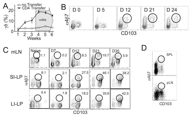
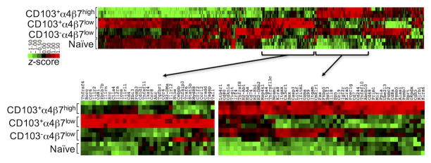
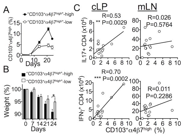
γδ T lymphocytes, dominant T cell subsets in the intestine, mediate both regulatory and pathogenic roles, yet the mechanisms underlying such opposing effects remain unclear. In this study, we identified a unique γδ T cell subset that coexpresses high levels of gut-homing integrins, CD103 and α4β7. They were exclusively found in the mesenteric lymph node after T cell-mediated colitis induction, and their appearance preceded the inflammation. Adoptive transfer of the CD103+α4β7high subsets enhanced Th1/Th17 T cell generation and accumulation in the intestine, and the disease severity. The level of generation correlated with the disease severity. Moreover, these cells were also found to be elevated in a spontaneous mouse model of ileitis. Based on the procolitogenic function, we referred to this subset as "inflammatory" γδ T cells. Targeting inflammatory γδ T cells may open a novel strategy to treat inflammatory diseases where γδ T cells play a pathogenic role including inflammatory bowel disease.


Figures

Figure 1
Figure 1. Gut homing integrin expression of γδ T cells in steady state conditions
(A) α4β7 and CD103 expression on γδ T cells or CD4 T cells is shown. pLN, peripheral lymph node. mLN, mesenteric lymph node. LP, lamina propria. IEL, intraepithelial lymphocyte. (B) mLN, LP, IEL, and pLN cells from WT or TCRβ−/− mice were stained for γδ TCR, α4β7, and CD103. Integrin expression of mLN γδ T cells are shown in grey zebra plots. Overlaid dot plots represent integrin expression of LP, IEL, and pLN γδ T cells. (C) CD69 expression of mLN, SI-LP, and SI-IEL γδ T cells. Each experiment was carried out at least three times, with results similar to the representative examples shown.
Figure 2
Figure 2. CD103+α4β7high γδ T cell subsets during colitic inflammation
(A) Groups of TCRβ−/− mice were induced for colitis by transferring naïve CD4 T cells. The mice were weekly bled and stained for γδ T cells. Data shown are the mean ± SD of 5 mice. (B) TCRβ−/− mice were transferred with naïve CD4 T cells. α4β7 and CD103 expression of circulating γδ T cells was determined by flow analysis. (C) γδ T cell integrin expression in the indicated tissues was determined following CD4 T cell transfer. (D) Integrin expression of γδ T cells in SPL and pLN was measured 3 weeks after CD4 T cells transfer. Each experiment was carried out at least three times, with results similar to the representative examples shown.
Figure 3
Figure 3. Gut homing expression on γδ T cells was dependent on CD4 T mediated inflammation
Naïve CD4 T cells and lymphoid γδ T cells were cotransferred into Rag1−/− mice. The mice were sacrificed 3 weeks post transfer, and γδ T cell expression of α4β7 and CD103 was determined by flow analysis. Data shown are representative of three independent experiments.
Figure 4
Figure 4. Characterization of γδ T cell subsets
(A) TCRβ−/− recipients of naïve CD4 T cells were sacrificed 3 weeks post transfer, and γδ T cells in the mLN, SI, and large intestine (LI) were examined for Ki67 expression. Based on the CD103 and a4b7 expression, each subset was numbered. (B) Vγ4 expression on each γδ T cell subset was measured. (C) Vγ1 and Vγ7 expression of the indicated mLN γδ T cell subsets were also measured. Data shown are the mean ± SD of 5–7 individually tested mice from two independent experiments. Vγ expression is a representative of two or three independent experiments. ***, p<0.001; ****, p<0.0001.
Figure 5
Figure 5. Gene expression profiles in different γδ T cell subsets
Different γδ T cell subsets (n=3, CD103+α4β7high, CD103+α4β7low, and CD103α4β7low) were FACS sorted from the mLN of TCRβ−/− recipients after 3 weeks post naive CD4 T cell transfer. Total γδ T cells from naïve TCRβ−/− mice were also isolated and used as a control. RNA expression was compared by a Nanostring analysis.
Figure 6
Figure 6. CD103+α4β7high cells are phenotypically distinct
(A) TCRβ−/− mice were transferred with naïve CD4 T cells and sacrificed 3 weeks post the transfer. γδ T cells in the mLN and IEL were examined for CD8α and CD8β expression by flow analysis. Naive TCRβ−/− mice were included as a non-inflammatory control. (B and C) mLN (B) and IEL (C) cells were harvested 3 weeks post colitis induction, and stimulated with PMA/Ionomycin for intracellular cytokine production. The data shown are representative of two independent experiments.
Figure 7
Figure 7. CD103+α4β7high mLN γδ T cells induce severe colitis following transfer
(A) CD103+α4β7high and CD103+α4β7low γδ T cells were FACS sorted from mLN and LP of TCRβ−/− mice that received naïve CD4 T cells 3 weeks earlier, and transferred into Rag1−/− mice together with naïve CD4 T cells. Weight loss was weekly monitored. (B) H&E staining of the colon tissues. (C) Histology score. (D and E) The recipients were sacrificed 5 weeks post transfer, and donor CD4 T cell accumulation in the mLN (D) and colon (E) was determined. Cells were also ex vivo stimulated for intracellular IFNγ and IL-17 expression. (F) CD103+α4β7high and CD103+α4β7low subsets derived from TcrdGFP TCRβ−/− mice were transferred into Rag1−/− mice together with naïve CD4 T cells. GFP+ γδ T cell accumulation in the LI-LP and mLN was determined 5 weeks post transfer. Data shown are the mean ± SD of 6–8 individually tested mice from two independent experiments. *, p<0.05; **, p<0.01; ***, p<0.001.
Figure 8
Figure 8. The level of circulating CD103+α4β7high γδ T cell subsets correlates with disease severity
(A) TCRβ−/− mice (n=18) received naïve CD4 T cells, and blood CD103+α4β7high γδ T cells were monitored weekly. CD103+α4β7high γδ-high and CD103+α4β7high γδ-low groups were divided based on the level of circulating CD103+α4β7high γδ T cell levels. The mean SD of CD103+α4β7high γδ levels is shown. (B) Body weight loss from each group was compared. (C) The mice were sacrificed 25 days post transfer. Circulating CD103+α4β7high γδ T cells were measured. mLN and LP cells were ex vivo stimulated to measure IFNγ- and IL-17-producing CD4 T cells. Blood CD103+α4β7high γδ T cell levels were compared to cytokine producing CD4 T cells in the tissues. *, p<0.05.
Figure 9
Figure 9. CD103+α4β7high γδ T cell levels in mice with spontaneous intestinal inflammation
SAMP1/YitFc mice and age-matched AKR control mice were examined for CD103+α4β7high γδ T cells in the mLN and LI-LP tissues. Data shown are the mean ± SD of 4–5 individually tested animals. *, p<0.05; **, p<0.01; ***, p<0.001.

References

    1. Carding SR, Egan PJ. Gammadelta T cells: functional plasticity and heterogeneity. Nat Rev Immunol. 2002;2:336–345. - PubMed
    1. Chennupati V, Worbs T, Liu X, Malinarich FH, Schmitz S, Haas JD, Malissen B, Forster R, Prinz I. Intra- and intercompartmental movement of gammadelta T cells: intestinal intraepithelial and peripheral gammadelta T cells represent exclusive nonoverlapping populations with distinct migration characteristics. Journal of immunology. 2010;185:5160–5168. - PubMed
    1. Edelblum KL, Shen L, Weber CR, Marchiando AM, Clay BS, Wang Y, Prinz I, Malissen B, Sperling AI, Turner JR. Dynamic migration of gammadelta intraepithelial lymphocytes requires occludin. Proceedings of the National Academy of Sciences of the United States of America. 2012;109:7097–7102. - PMC - PubMed
    1. Hayday A, Tigelaar R. Immunoregulation in the tissues by gammadelta T cells. Nat Rev Immunol. 2003;3:233–242. - PubMed
    1. Ribot JC, deBarros A, Pang DJ, Neves JF, Peperzak V, Roberts SJ, Girardi M, Borst J, Hayday AC, Pennington DJ, Silva-Santos B. CD27 is a thymic determinant of the balance between interferon-gamma- and interleukin 17-producing gammadelta T cell subsets. Nature immunology. 2009;10:427–436. - PMC - PubMed

Publication types