

Case Reports
doi: 10.4269/ajtmh.15-0902.
Epub 2016 Oct 17.
Cerebral Paragonimiasis Presenting with Sudden Death
Affiliations
- PMID: 27928089
- PMCID: PMC5154461
- DOI: 10.4269/ajtmh.15-0902
Item in Clipboard
Case Reports
Cerebral Paragonimiasis Presenting with Sudden Death
Am J Trop Med Hyg.
.
Abstract
A 58-year-old Korean-born woman with a history of seizures and psychiatric issues was found dead at home. Autopsy was notable for large, calcified nodules that had nearly replaced her right temporal lobe. Histologic examination revealed the presence of Paragonimus eggs. This case demonstrates a rare manifestation of an aberrantly migrated lung fluke that resulted in epilepsy and sudden death years after the initial infection.
© The American Society of Tropical Medicine and Hygiene.
Figures
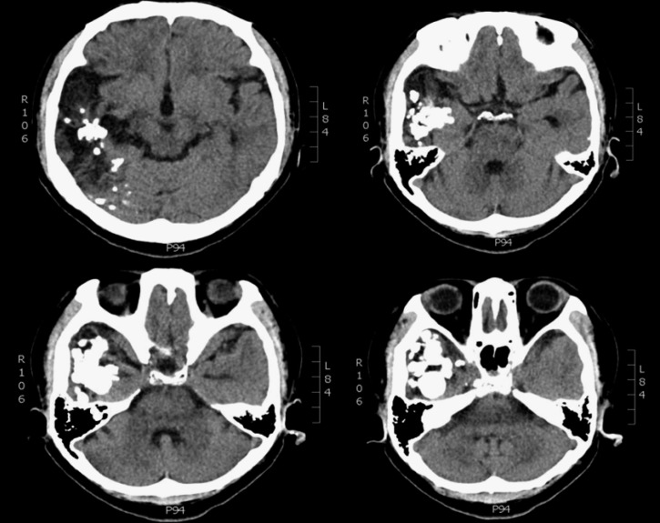
Axial images demonstrate a large area of hypoattenuation and dystrophic calcification involving the right lateral temporal lobe, right inferior parietal lobe, and right lateral occipital lobe.

Coronal sections of formalin-fixed whole brain with yellow discoloration and induration of right temporal lobe.
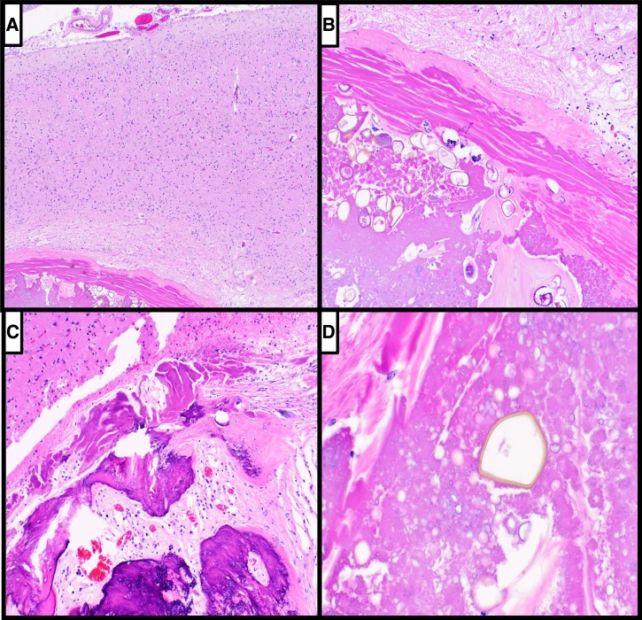
(A) Hematoxylin and eosin–stained sections of formalin-fixed brain tissue reveal a gliotic rim of temporal lobe around walled-off, necrotic, and partially calcified debris (×40). (B) Higher magnification reveals abundant necrotic debris and numerous intermingled eggs (×100). (C) Focally, osseous metaplasia, testament to the long-standing nature of the lesion, is identified (×100). (D) Careful scrutiny of the parasitic egg reveals a thick, golden-brown hue of the egg wall and lack of spine, consistent with Paragonimus (×400).
References
-
- Chen J, Chen Z, Lin J, Zhu G, Meng H, Cui G, Wu N, Hu R, Pan J, Zou Y, Feng H. Cerebral paragonimiasis: a retrospective analysis of 89 cases. Clin Neurol Neurosurg. 2013;115:546–551. - PubMed
-
- Xia Y, Ju Y, Chen J, You C. Cerebral paragonimiasis: a retrospective analysis of 27 cases. J Neurosurg Pediatr. 2015;15:101–106. - PubMed
-
- Oh SJ. Cerebral paragonimiasis. J Neurol Sci. 1969;8:27–48. - PubMed
-
- Kusner DJ, King CH. Cerebral paragonimiasis. Semin Neurol. 1993;13:201–208. - PubMed
-
- Chai JY. Paragonimiasis. Handb Clin Neurol. 2013;114:283–296. - PubMed
Publication types
MeSH terms
Grants and funding
LinkOut - more resources
Full Text Sources
Other Literature Sources
Medical
