

Histopathological Changes of Inner Retina, Optic Disc, and Optic Nerve in Rabbit with Advanced Retinitis Pigmentosa
- PMID: 27928420
- PMCID: PMC5120756
- DOI: 10.1080/01658107.2016.1229339
Histopathological Changes of Inner Retina, Optic Disc, and Optic Nerve in Rabbit with Advanced Retinitis Pigmentosa
Abstract
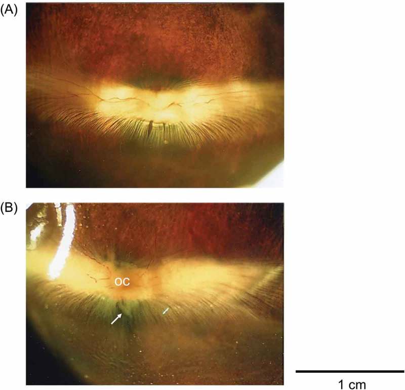
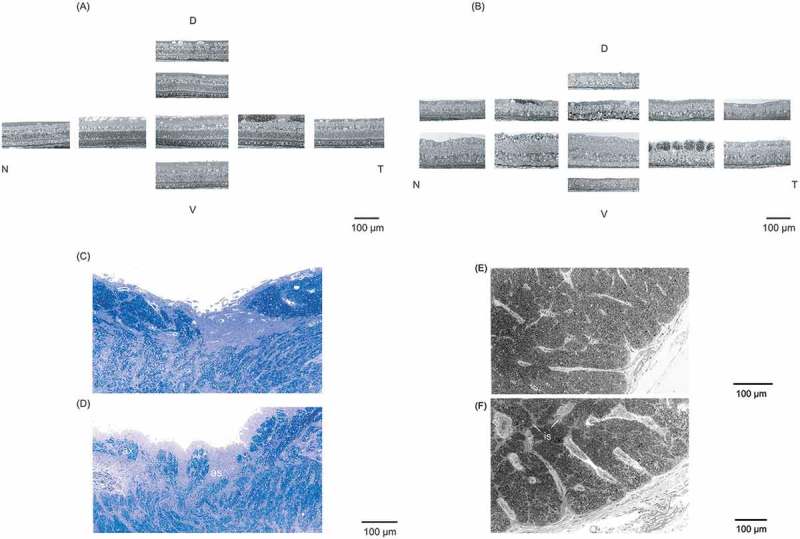
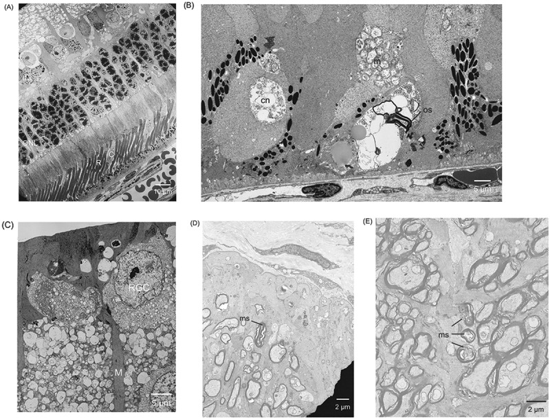
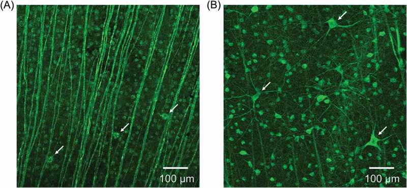
We observed the histopathological changes of retinal ganglion cells (RGCs), optic disc, and optic nerve in rabbit with advanced retinitis pigmentosa (RP). Wild-type (WT) and rhodopsin transgenic (Tg) of RP rabbits were used at age 24 months. Light and electron microscopy were used to observe the retina, optic disc, and optic nerve. RGCs were also confirmed by immunofluorescent staining with a TUJ-1 monoclonal antibody. In addition to the rod and cone degeneration, we observed the astrocyte infiltration of the optic disc due to the damage of small RGCs and nerve fibres and atrophy of small optic nerve fibres. They subsequently lead to the optic disc excavation and atrophy of the optic nerve. Consequently, our histopathological study clarified that not only the outer retina but also the inner retina, the optic disc, and the optic nerve were also affected in the late stages of RP rabbit.
Keywords: Optic disc; optic nerve; rabbit; retinal ganglion cell; retinitis pigmentosa.
Figures
References
-
- Berson EL. Retinitis pigmentosa. The Friedenwald Lecture. Invest Ophthalmol Vis Sci 1993;34:1659–1676. - PubMed
-
- Hartong DT, Berson EL, Dryja TP.. Retinitis pigmentosa. Lancet 2006;368:1795–1809. - PubMed
-
- Kondo M, Sakai T, Komeima K, Kurimoto Y, Ueno S, Nishizawa Y, Usukura J, Fujikado T, Tano Y, Terasaki H.. Generation of a transgenic rabbit model of retinal degeneration. Invest Ophthalmol Vis Sci 2009;50:1371–1377. - PubMed
LinkOut - more resources
Full Text Sources
Other Literature Sources
Miscellaneous