. 2016:2:16019.
doi: 10.1038/npjamd.2016.19. Epub 2016 Sep 22.

Extracellular Vesicle-Associated Aβ Mediates Trans-Neuronal Bioenergetic and Ca2+-Handling Deficits in Alzheimer's Disease Models

Affiliations

Extracellular Vesicle-Associated Aβ Mediates Trans-Neuronal Bioenergetic and Ca2+-Handling Deficits in Alzheimer's Disease Models

Erez Eitan et al. NPJ Aging Mech Dis. 2016.

Abstract

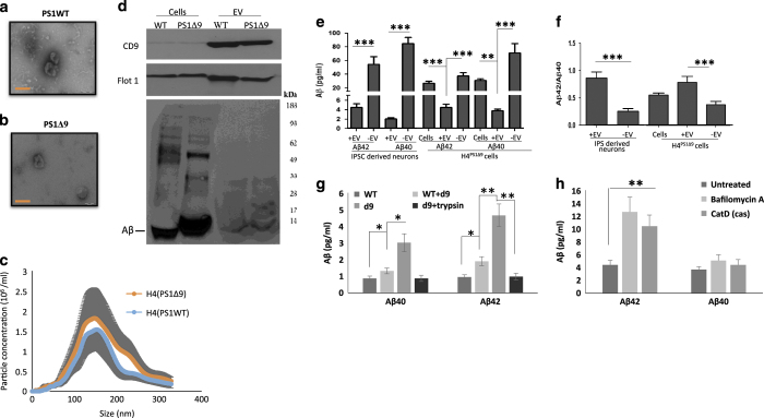
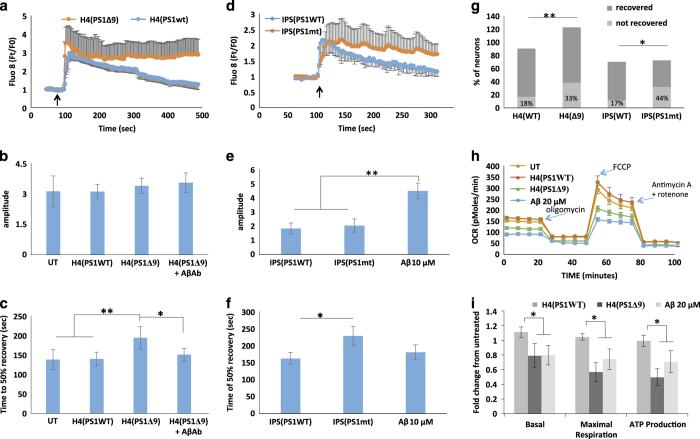
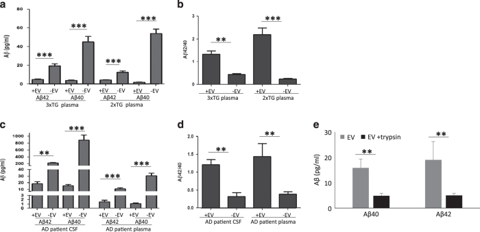
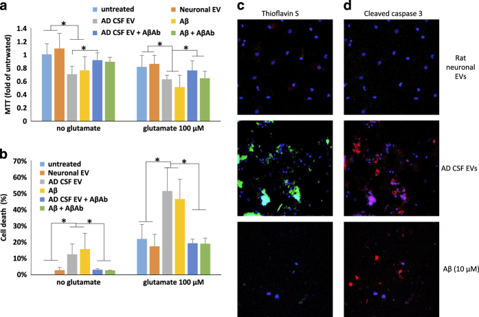
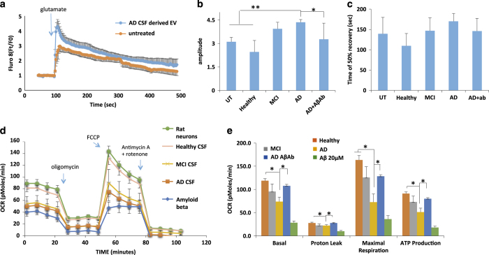
Alzheimer's Disease (AD) is an age-related neurodegenerative disorder in which aggregation-prone neurotoxic amyloid β-peptide (Aβ) accumulates in the brain. Extracellular vesicles (EVs) are small 50-150 nanometer membrane vesicles that have recently been implicated in the prion-like spread of self-aggregating proteins. Here we report that EVs isolated from AD patient CSF and plasma, from the plasma of two AD mouse models, and from the medium of neural cells expressing familial AD presenilin 1 mutations, destabilize neuronal Ca2+ homeostasis, impair mitochondrial function, and sensitize neurons to excitotoxicity. EVs contain a relatively low amount of Aβ but have an increased Aβ42/ Aβ40 ratio; the majority of Aβ is located on the surface of the EVs. Impairment of lysosome function results in increased generation EVs with elevated Aβ42 levels. EVs may mediate transcellular spread of pathogenic Aβ species and that impair neuronal Ca2+ handling and mitochondrial function, and may thereby render neurons vulnerable to excitotoxicity.

Keywords: Alzheimer's disease; amyloid β-peptide; exosomes; extracellular vesicles; glutamate; mitochondria.


Conflict of interest statement

The authors declare no conflict of interest.

Figures

Figure 1
Figure 1
Human neural cells expressing familial Alzheimer’s disease (AD) presenilin 1 mutations release extracellular vesicles (EVs) with elevated levels of Aβ42 on their outer surface. (a, b). Transmission electron microscope images of EVs released from H4 cells expressing either the δ9 PS1 mutation (a) or wild-type (WT) PS1 (b) Bar = 100 nm. (c) Size distribution of EVs released from H4 cells expressing mutant (red) or wild type (blue) PS1 measured by Nanocyte particle tracking. (d) Immunoblot demonstrating enrichment of EV markers CD9 and Flotillin-1 in EVs released from H4 cells, and relatively low amounts of amyloid β-peptide (Aβ) in EVs relative to H4 cell lysate. (e, f) Levels of Aβ42 and Aβ40 (e) and the Aβ42/Aβ40 ratio (f) in cells, EVs and EV-depleted medium from neurons differentiated from induced pluripotent stem cell (iPSC) that were generated from fibroblasts from a patient with familial AD (PS1 mutation; n=5 cultures), and from H4 human neuroglioma cells expressing mutant PS1 (8 separate cultures). (g) Levels of Aβ40 and Aβ42 in EVs isolated from the medium of cultured H4Ps1Δ9 cells, H4Ps1WT cells, H4Ps1WT EVs incubated in H4Psn1Δ9 EV-depleted medium, or H4Psn1Δ9 EVs incubated with trypsin (n=3). (h) Levels of Aβ40 and Aβ42 in EVs isolated from the medium of cultured H4Psn1Δ9 cells that had been treated for 24 h with vehicle (control; n=5) or bafilomycin A (n=5), or in which cathepsin D was knocked down using CRISPR Cas9 technology (n=3). *P<0.05, **P<0.01, ***P<0.001.
Figure 2
Figure 2
Extracellular vesicles (EVs) isolated from the medium of cultured human neural cells expressing mutant PS1 are neurotoxic. (a, b) Rat primary cortical neurons were exposed to the indicated treatments for 24 h and cell viability was evaluated by MTT (a) and LDH (b) assays (n=3–6 separate cultures). (c) Thioflavin S (green), and 4′,6-diamidino-2-phenylindole dihydrochloride (DAPI; blue) staining demonstrates that treating cortical neuron cultures with Psn1Δ9-derived EVs results in the accumulation of Thioflavin S reactive aggregates. (d) Cleaved caspase 3 staining (green) and DAPI (blue) staining showing increased cell death following incubation of cortical neuron cultures with Psn1Δ9-derived but not WT EVs. (e). Reduced MAP2 staining (green; a neuronal protein) is observed in rat neuronal cultures incubated with Psn1Δ9-derived EVs. DAPI (blue). (f). Quantification of fluorescence intensities of primary neuronal cultures that had been exposed for 48 h to EVs derived from H4 cells expressing mutant PS1 and then stained with Thioflavin S, cleaved caspase 3 and MAP2. Values are expressed as fold difference from the value for neurons exposed to EVs from control H4 cells (n=6 cultures). (g) Images showing PKH-labeled EVs (red) and DAPI (blue) staining in cortical neurons that had been incubated for 6 h in the presence of EVs released from H4 cells expressing WT PS1 or mutant PS1 (PS1MT). (h, i). Levels of MTT reduction and LDH release (cytotoxicity) in cortical neurons that that had been incubated for 48 h in the presence of EVs from neurons derived from induced pluripotent stem cell (iPSC) generated from fibroblasts from a patient with familial Alzheimer’s disease (AD; IPS), EV-depleted iPSC culture medium, or Aβ1–42 (10 μmol/l). UT, untreated control cultures. Cultures were co-treated with glutamate (100 nmol/l) or vehicle as indicated. Values are the mean and s.e.m. (n=6 cultures). *P<0.05, **P<0.01. Aβ, amyloid β-peptide.
Figure 3
Figure 3
Extracellular vesicles (EVs) generated by human neural cells expressing mutant PS1 impair cellular Ca2+ handling and mitochondrial function in cerebral cortical neurons. (a) Preincubation of rat cortical neurons with H4Psn1Δ9-derived EVs for 24 h resulted in a greater sustained Ca2+ response to glutamate (10 μmol/l) stimulation (arrow indicates the time of glutamate application) compared with neurons preincubated with EVs from H4Psn1WT cells. Values are the mean and s.e.m. of measurements made in 20 neurons per culture in 7 different cultures. (b, c) Quantification of peak amplitude (b) and time to reach 50% recovery (c) following glutamate stimulation in cortical neurons that had been pretreated for 24 h with EVs (H4D9 cells with either wild type (WT) or Δ9 PS1; H4(PS1Δ9)+AβAb was a condition in which the EVs were incubated with an amyloid β-peptide (Aβ) antibody (6E10). Values are the mean and s.e.m. of measurements made in neurons from 7 different cultures. (d) Alzheimer’s disease (AD) patient iPSC-derived neuronal EVs induce Ca2+ dysregulation following stimulation with glutamate (10 μmol/l; arrow indicates the time of glutamate application). Values are the mean and s.e.m. of measurements made in three different cultures (20 neurons per culture). (e, f) Peak amplitude (e), and time to reach 50% recovery (f) following glutamate stimulation in rat cortical neurons pretreated for 24 h with EVs from AD patient or WT induced pluripotent stem cells (iPSC)-derived neurons (Aβ condition is rat cortical neurons incubated with 10 μmol/l synthetic Aβ1–42). (g). Analysis of the percentage of cortical neurons unresponsive to glutamate following incubation with mutant or wild-type (WT) EVs derived from H4 cells and IPSC-derived neurons (n=7 for H4 EVs treated neurons and n=3 for IPC EVs treated neurons). (h) Representative Seahorse oxygen consumption assay showing mitochondrial impairment 24 h following treatment with H4Psn1Δ9-derived EVs or 20 μmol/l Aβ1–42. (i) Basal oxygen-consumption rate, maximal consumption rate and ATP production in cortical neurons that had been treated for 24 h with H4Psn1Δ9-derived EVs or 20 μmol/l Aβ1–42. Values are the mean and s.e.m. from measurements made in five independent cultures. *P<0.05, **P<0.01.
Figure 4
Figure 4
Extracellular vesicles (EVs) isolated from cerebropspinal fluid (CSF) and plasma of Alzheimer’s disease (AD) patients, and from plasma in two AD mouse models, exhibit a high Aβ42/40 ratio. Concentrations of Aβ40 and Aβ42 were measured in samples of EVs (+EV) and corresponding fluids from which they were isolated (−EV). (a, b). Results of analyses of blood plasma samples from 3xTgAD and 2xTgAD mice (n=5 3xTgAD and n=6 2xTgAD mice). (c, d). Results of analyses of CSF and blood plasma samples from AD patients (n=6 patients). (e) Quantification of Aβ40 and Aβ42 levels in EVs isolated from the CSF of AD patients. The EVs were incubated with or without with trypsin prior to the analysis (EV preparations from three different AD patients). **P<0.01, ***P<0.001. Aβ, amyloid β-peptide.
Figure 5
Figure 5
Alzheimer’s disease (AD) patient CSF-derived extracellular vesicles (EVs) are neurotoxic. (a, b) Results of MTT and LDH assays demonstrating that 48 h incubation of rat neurons with AD cerebrospinal fluid (CSF) EVs (at a concentration of 100 EVs per neuron) diminished neuronal survival by an amount similar to that of neurons exposed to Aβ1–42 (10 μmol/l). AD CSF EVs also increased neuronal vulnerability to glutamate during a 24 h incubation (n=6 cultures). Where indicated, cultures were co-treated with Aβ antibody 6E10 (1 μg antibody per 107 EVs). (c, d) Cultured cortical neurons were incubated for 24 h in the presence of fluorescently tagged EVs that had been isolated from the culture medium of rat cortical neurons or from AD CSF (100 EVs per neuron), or with 10 μmol/l Aβ1–42. Cortical neurons were then stained with Thioflavin S or anti-cleaved caspase 3. Representative images are shown. Similar results were obtained in six separate experiments. *P<0.05. Aβ, amyloid β-peptide.
Figure 6
Figure 6
Alzheimer’s disease (AD) patient cerebrospinal fluid (CSF)-derived extracellular vesicles (EVs) impair neuronal Ca2+ regulation and mitochondrial function. (a) Graph showing relative intracellular Ca2+ levels (Fluo 8 fluorescence intensity before and during exposure to glutamate (100 μmol/l). (b, c) Graphs showing amplitudes of peak Ca2+ responses to glutamate (b) and the time required for the Fluo 8 fluorescence intensity to recover to 50% of the peak level (c). (d, e) Results of Seahorse analysis of mitochondrial respiration in cultured neurons that had been pretreated for 24 h with EVs isolated from the medium bathing healthy primary rat cortical neurons (100 particles per neuron), 10 μmol/l amyloid β-peptide (Aβ) or EVs isolated from the CSF of AD patients (100 particles per neuron). (d) Results of a representative experiment. (e) Data from six experiments. *P<0.05; **P<0.01.

References

    1. Mattson, M. P. Pathways towards and away from Alzheimer's disease. Nature 430, 631–639 (2004). - PMC - PubMed
    1. Karch, C. M. , Cruchaga, C. & Goate, A. M. Alzheimer's disease genetics: from the bench to the clinic. Neuron 83, 11–26 (2014). - PMC - PubMed
    1. Platt, T. L. , Reeves, V. L. & Murphy, M. P. Transgenic models of Alzheimer's disease: better utilization of existing models through viral transgenesis. Biochim. Biophys. Acta 1832, 1437–1448 (2013). - PMC - PubMed
    1. Bezprozvanny, I. & Mattson, M. P. Neuronal calcium mishandling and the pathogenesis of Alzheimer's disease. Trends Neurosci. 31, 454–463 (2008). - PMC - PubMed
    1. Stutzmann, G. E. & Mattson, M. P. Endoplasmic reticulum Ca(2+) handling in excitable cells in health and disease. Pharmacol. Rev. 63, 700–727 (2011). - PMC - PubMed