. 2017 Jul;254(4):1627-1637.
doi: 10.1007/s00709-016-1053-0. Epub 2016 Dec 7.

RNA processing body (P-body) dynamics in mesophyll protoplasts re-initiating cell division

Affiliations

RNA processing body (P-body) dynamics in mesophyll protoplasts re-initiating cell division

Dilbag S Bhullar et al. Protoplasma. 2017 Jul.

Abstract

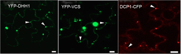
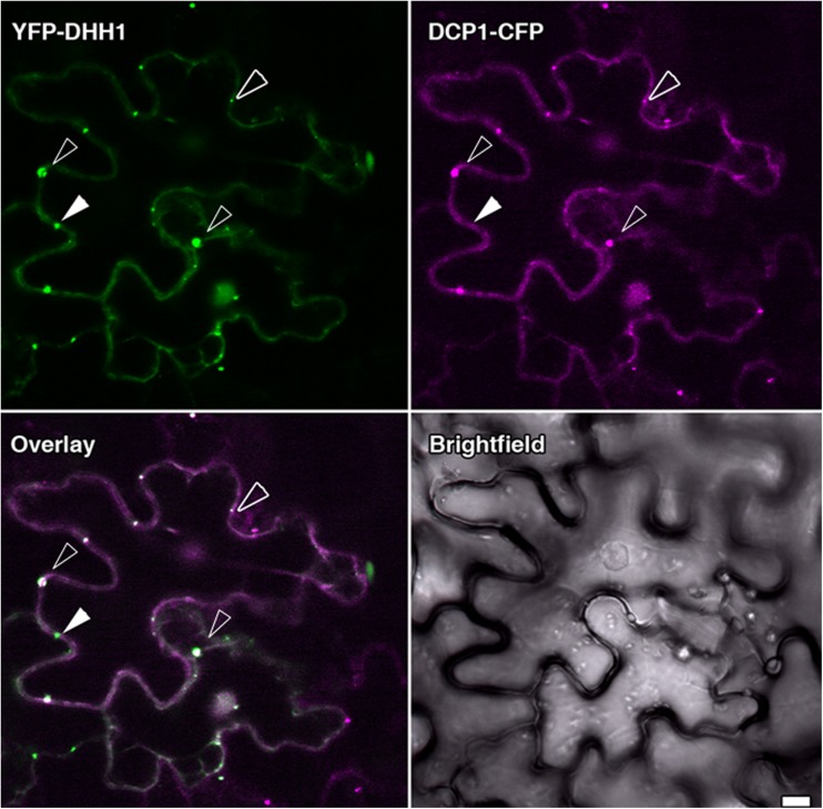
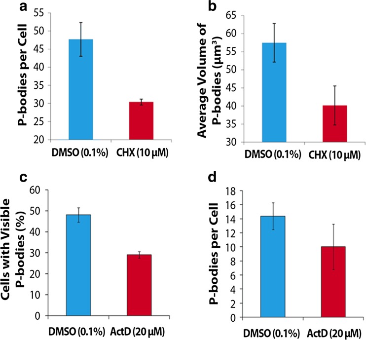
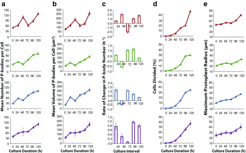
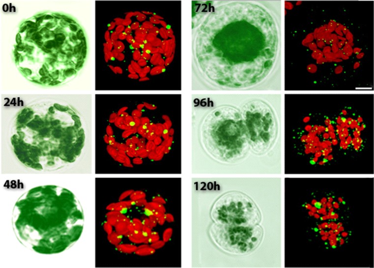
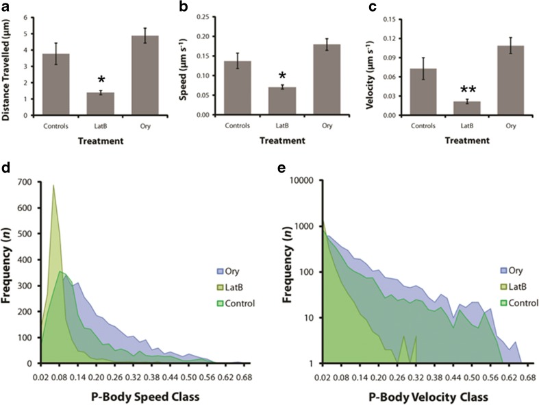
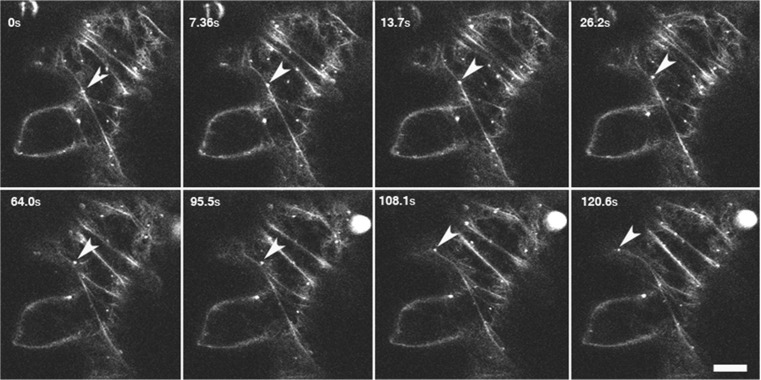
The ability of plants to regenerate lies in the capacity of differentiated cells to reprogram and re-enter the cell cycle. Reprogramming of cells requires changes in chromatin organisation and gene expression. However, there has been less focus on changes at the post transcription level. We have investigated P-bodies, sites of post transcriptional gene regulation, in plant cell reprogramming in cultured mesophyll protoplasts; by using a YFP-VARICOSE (YFP-VCSc) translational fusion. We showed an early increase in P-body number and volume, followed by a decline, then a subsequent continued increase in P-body number and volume as cell division was initiated and cell proliferation continued. We infer that plant P-bodies have a role to play in reprogramming the mature cell and re-initiating the cell division cycle. The timing of the first phase is consistent with the degredation of messages no longer required, as the cell transits to the division state, and may also be linked to the stress response associated with division induction in cultured cells. The subsequent increase in P-body formation, with partitioning to the daughter cells during the division process, suggests a role in the cell cycle and its re-initiation in daughter cells. P-bodies were shown to be mobile in the cytoplasm and show actin-based motility which facilitates their post-transcriptional role and partitioning to daughter cells.

Keywords: Dedifferentiation; P-bodies; Protoplast division; RNA decapping; RNA degredation; RNA storage.


Conflict of interest statement

The authors have no conflict of interest.

Figures

Fig. 1
Fig. 1
Subcellular localisation of DHH1, VCSc and DCP1 in tobacco epidermal leaf cells. Proteins are labelled with YFP (VCSc and DHH1; pseudo-coloured green) or CFP (DCP1; pseudo-coloured red). White arrows indicate P-bodies. P-bodies could be visualised as bright cytoplasmic dots in YFP-DHH1, YFP-VCSc and DCP1- CFP. Single focal plane images are shown. Bars =20 μm
Fig. 2
Fig. 2
Co-localization analysis of YFP-DHH1 with DCP1- CFP. DCP1- CFP was co-expressed with YFP-DHH1 in tobacco epidermal leaf cells. Blank arrowheads indicate the cytoplasmic foci marked by both proteins. While DCP1 frequently localised to cytoplasmic foci marked by DHH1, at times DHH1 marked P-bodies devoid of DCP1 were observed as well. Solid white arrows indicate cytoplasmic foci marked by DHH1 lacking DCP1. Bars =10 μm
Fig. 3
Fig. 3
Quantitative analysis of P-body dynamics in CHX and ActD treated protoplasts. (a) P-body number per cell was significantly reduced by 10 μM CHX, p = 0.008, and (b) volume was reduced, p = 0.081. (c) In samples treated with 20 μM ActD a significant reduction in the number of cells with visible P-bodies was observed, p = 0.010, and (d) there was a smaller reduction in P-body numbers, p = 0.380. A students’ t-test was applied to each data set and p-values are given. Protoplasts were from transiently transformed leaves and the marker fusion protein was YFP-VCSc
Fig. 4
Fig. 4
P-body dynamics during mesophyll protoplast dedifferentiation and re-entry into the cell cycle. Replicate one shown in red, two in green, three in blue and the average in purple. (a) The number of P-bodies observed in cultured mesophyll protoplasts. Note that all replicates show a similar trend, that is two phases of P-body proliferation, however, replicates two and three are shifted to earlier culture changes relative to replicate one, leading to a masking of the biphasic proliferation trend in the averaged results (purple trace). (b) Total volume of P-bodies in a cell. Similar trend as to that shown in (a) and consistent with little change in the average size of P-bodies over the culture interval. (c) Rates of change in P-body number. Note that on average (purple graph), the biphasic burst of P-body proliferation is clearly evident. (d) Proportion of cells divided. On average (purple trace), cell division increased linearly after 48 h culture. Note that the increase in P-body number appears to correlate with the amount of cell division. (e) The maximum protoplasts radius, an indicator of cell volume and therefore cell expansion during culture. Error bars represent SE. Protoplasts were isolated from tobacco leaves from transgenic plants expressing YFC-VCSc
Fig. 5
Fig. 5
Visual observations of cultured protoplasts. Protoplasts isolated from transgenic tobacco leaves expressing YFC-VCSc observed over 120 h. Images taken at 24 h time intervals from 0 h (isolation time point) to 120 h (cultured protoplasts). Simultaneous visualisation of autofluoroscent chloroplasts provides insights into cell division pattern. Chloroplasts (red) are distributed across the entire cell area (0–48 h), however they cluster around the nucleus (72 h) before the onset of cell division, as earlier described (Sheahan et al. 2004a). Cell division shown at 96 h and 120 h. P-bodies (small green spheres) can be observed distributed throughout the cell with no particular localization. It appears that P-bodies are increasing in number as the culture progresses (0–120 h). The images of representative protoplasts at the different time points show a general trend of cell division, increased P-body number and chloroplast clustering. Bar: 10 μm
Fig. 6
Fig. 6
Tracking of P-body movements showing motion paths of P-bodies. Transiently transformed tobacco epidermal leaf cells were visualised for 200 s. P-bodies generally exhibited erratic and circular motion (a,b) while occasional rapid vectorial movements were also observed (c). Bars =10 μm
Fig. 7
Fig. 7
(a-c) Means of five replicates with SE for time-lapse image series depicting the effects of LatB and Ory on P-body motility (a), speed (b) and velocity (c) in tobacco epidermal leaf cells. LatB treatment reduced vectorial movements of P-bodies, best represented by the velocity analysis (c), while speed was also reduced to a lesser extent (b). (d-e) Frequency area graphs summarising the motile characteristics for the whole population of P-bodies analysed in each treatment. Most P-bodies exhibited lower speeds (d) and velocities (e) consistent with a predominantly erratic motility. Treatment with LatB however, skewed the results toward the left of the graph with substantially lower speeds, and more significantly, much lower velocities. Note that (e) has a logarithmic ordinate axis. Speed is distance/unit time (μm s−1) while velocity is displacement/unit time (μm s−1). P-bodies and AFs were visualised simultaneously in tobacco epidermal cells co-transfected with YFP-VCSc (P-body marker) and GFP-faBD2 (AF label). Standard errors indicated and * p < 0.05, ** p < 0.01 for a, b and c LatB versus control. Ory in all cases not significantly different to control
Fig. 8
Fig. 8
P-body actin colocalisation. The actin cytoskeleton can be observed as fine filaments and P-bodies as round white spheres in tobacco epidermal leaf cells. Images series was taken over a period of 2 min. Arrows represent a P-body that appears to travel on an AF bundle. P-bodies and AFs were visualised simultaneously in tobacco epidermal cells co-transfected with YFP-VCSc (P-body marker) and GFP-faBD2 (AF label)

References

    1. Abràmoff MD, Magalhães PJ, Ram SJ. Image processing with ImageJ. Biophotonics Intern. 2004;11:36–42.
    1. Aizer A, Brody Y, Ler LW, Sonenberg N, Singer RH, Shav-Tal Y. The dynamics of mammalian P body transport, assembly, and disassembly in vivo. Mol Biol Cell. 2008;19:4154–4166. doi: 10.1091/mbc.E08-05-0513. - DOI - PMC - PubMed
    1. Anderson P, Kedersha N. RNA granules. J. Cell Biol. 2006;172:803–808. doi: 10.1083/jcb.200512082. - DOI - PMC - PubMed
    1. Balagopal V, Parker R. Polysomes, P bodies and stress granules: states and fates of eukaryotic mRNAs. Curr Opin Cell Biol. 2009;21:403–408. doi: 10.1016/j.ceb.2009.03.005. - DOI - PMC - PubMed
    1. Brengues M, Teixeira D, Parker R. Movement of eukaryotic mRNAs between polysomes and cytoplasmic processing bodies. Science. 2005;310:486–489. doi: 10.1126/science.1115791. - DOI - PMC - PubMed