. 2016 Dec 8:7:13739.
doi: 10.1038/ncomms13739.

Export of piRNA precursors by EJC triggers assembly of cytoplasmic Yb-body in Drosophila

Affiliations

Export of piRNA precursors by EJC triggers assembly of cytoplasmic Yb-body in Drosophila

Cynthia Dennis et al. Nat Commun. .

Abstract

PIWI-interacting RNAs (piRNAs) are effectors of transposable element (TE) silencing in the reproductive apparatus. In Drosophila ovarian somatic cells, piRNAs arise from longer single-stranded RNA precursors that are processed in the cytoplasm presumably within the Yb-bodies. piRNA precursors encoded by the flamenco (flam) piRNA cluster accumulate in a single focus away from their sites of transcription. In this study, we identify the exportin complex containing Nxf1 and Nxt1 as required for flam precursor nuclear export. Together with components of the exon junction complex (EJC), it is necessary for the efficient transfer of flam precursors away from their site of transcription. Indeed, depletion of these components greatly affects flam intra-nuclear transit. Moreover, we show that Yb-body assembly is dependent on the nucleo-cytoplasmic export of flam transcripts. These results suggest that somatic piRNA precursors are thus required for the assembly of the cytoplasmic transposon silencing machinery.


Figures

Figure 1
Figure 1. Sub-cellular localization of flam piRNA precursor transcripts in ovarian follicle cells.
Examples of subcellular localization of flam precursors in ovarian follicle cells. flam transcripts revealed by flam 508 RNA probe are in red and nuclear periphery revealed with anti-lamin antibody is visualized in green.
Figure 2
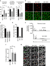
Figure 2. Exportin Nxt1 and Nxf1 are involved in flam RNA export and intra-nuclear traffic to Dot COM.
(a) Quantitative analysis of flam transcript localization in ovarian follicle cells. The percentage of cells with a nuclear (left), transmembrane (middle) and cytoplasmic (right) focus is plotted. Error bars represent s.e.m. n=45 (ISO1A), n=28 (Nxt1), n=22 (Nxf1), n is the number of independent experiments. ****P value <0.0001 and **P value <0.01 are calculated according to Mann–Whitney test. (b) flam transcripts (red) and nuclear membrane (green) are visualized using flam 508 RNA probe and anti-lamin antibody, respectively. One Z-stack of confocal image is shown. Therefore, as flam foci are not all in the same focus they cannot be all visualized in every nucleus of the field. (c) Fold change in steady-state RNA level of flam in Nxt1-SKD ovaries compared with ISO1A ovaries. RNA levels are normalized to rp49 level. The positions of the primers used are indicated above and their sequence is given in Supplementary Table 2. Data are presented as means (n=3). Error bars indicate s.e.m. *P value <0.05, according to the Student's t-test. (d,e) Changes in steady-state levels of flam unique 23–29 nt piRNAs in ovarian cells of ISO1A and Nxt1- and Nxf1-SKD lines measured by small RNA sequencing (normalized to one million genome-mappable reads). (d) Density plot of unique 23–29 nt piRNAs mapping to the flam piRNA cluster. The Y and X axes in e are identical for the three graphs. X axis represents the flam 180 kb starting from its transcription start site on the left. Y axis represents the quantity of piRNAs normalized to one million genome mappable reads. (f) Quantitative analysis of relative localization between flam DNA and flam transcripts in follicle cells. The percentage of cells with a DNA–RNA co-localization is plotted. Error bars represent s.e.m. n=32 (ISO1A), n=20 (Nxt1), n=38 (Nxf1), n is the number of independent experiments. ****P value<0.0001, ***P value <0.001), according to Mann–Whitney test. Error bars represent s.d. *P value <0.05, according to Mann–Whitney test. (g) Double DNA/RNA FISH experiments. flam DNA (green) and flam transcripts (red) are respectively detected with DNA probe and 508 riboprobe. DNA is stained with Hoescht (white).
Figure 3
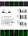
Figure 3. flam transcripts are correctly spliced and exported in EJC- and UAP56-depleted lines.
(a) Piwi (magenta) and nuclear membrane (green) are visualized by immunofluorescence using anti-Piwi and anti-lamin antibodies, respectively, in ISO1A and Mago-, RnpS1-, Tsu-, Acn- and UAP56-SKD lines. (b) Genomic structure of flam piRNA cluster 5′ end showing donor (position +432, +583, +748) and acceptor (position +2,068) splicing sites regarding +1 position of flam TSS (black arrow). Alternative splicing of intron 1 is shown above by the grey lines. Grey arrows indicate the position of primers used for RT–PCR whose sequence is given in Methods section. (c) RT–PCR on WT (w1118) genomic DNA and cDNAs from w1118-, Mago-, RnpS1-, Tsu- and Acn-SKD ovaries are obtained after DNAse I treatment of total ovarian extracted RNAs followed (+RT) or not (−RT) by reverse transcription. Amplification of unspliced molecule gives rise to 2,146 bp DNA (lane 1). Spliced RNAs resulting from first, second or third donor splicing sites give rise to 827, 662, 511 bp fragments (lanes 3, 5, 7, 9, 11). Black arrowheads indicate the position of specific amplified DNA. (d) flam transcripts (red) and nuclear membrane (green) are visualized by RNA-FISH coupled to immunofluorescence using flam 508 RNA probe and anti-lamin antibody, respectively, in ovarian follicle cells of WT ISO1A and Mago-, RnpS1-, Tsu-, Acn- and UAP56-SKD lines. (e) Quantitative analysis of flam transcript localization in ovarian follicle cells of these depleted lines. The percentage of cells with nuclear, transmembrane or cytoplasmic foci is plotted. Error bars represent s.e.m. n=13 (ISO1A), n=13 (Mago), n=14 (Rnps1), n=12 (Tsu), n=16 (Acn), n=12 (UAP56), n is the number of independent experiments.
Figure 4
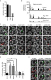
Figure 4. Components of EJC and UAP56 protein are required for flam transcript intra-nuclear traffic to Dot COM.
(a,b) Changes in steady-state levels of flam-unique 23–29 nt piRNAs in ovarian cells of ISO1A and Mago- and RnpS1-SKD lines measured by small RNA sequencing (normalized to one million genome-mappable reads). (a) Density plot of unique 23–29 nt piRNAs mapping to the flam piRNA cluster. The Y and X axes in b are identical for the three graphs. X axis represents the flam 180 kb starting from its transcription start site on the left. Y axis represents the quantity of piRNAs normalized to one million genome mappable reads. (c,e) Double DNA/RNA FISH experiments staining flam DNA (green) and flam transcripts (red) in ovarian follicle cells of WT ISO1A and Mago-, RnpS1-, Tsu-, Acn- and UAP56-SKD lines (c) or of Tsu-, Acn- and UAP56-depleted lines (e). DNA is stained with Hoescht (white). (d) Quantitative analysis of relative localization between flam DNA and flam transcripts in ovarian follicle cells. The percentage of cells with a DNA–RNA co-localization is plotted. Error bars represent s.e.m. n=32 (ISO1A), n=40 (Mago), n=26 (Rnps1), n=34 (Tsu), n=34 (Acn), n=18 (UAP56), n is the number of independent experiments. ****P value <0.0001, ***P value <0.001, *P value <0.05, according to Mann–Whitney test.
Figure 5
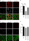
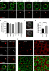
Figure 5. Yb-body formation is altered in Nxt1- and Nxf1- but not in EJC-depleted follicle cells.
(a,c) Armi (red), Yb (white) and nuclear membrane (green) are visualized by immunofluorescence in ISO1A and Nxt1- and Nxf1-SKD follicle cells (a), and in ISO1A and Mago- and RnpS1-depleted cells (c). (b,d) Quantitative analysis of Armi foci in ISO1A (n=16), Nxt1-(n=19) and Nxf1-SKD (n=16) follicle cells (b) and in ISO1A (n=16) and Mago-(n=15) and RnpS1-depleted cells (n=15), n is the number of independent experiments (d). The percentage of cells harbouring one or more Armi foci is plotted. Error bars represent s.e.m., ***P value <0.001, according to Mann–Whitney test.
Figure 6
Figure 6. Yb-bodies are assembled at the site of export of flam transcripts and fail to form foci in absence of flam transcripts
(a,e) flam transcripts (red), Armi foci (white) and nuclear membrane (green) are visualized by RNA-FISH coupled to immunofluorescence in ovarian follicle cells of ISO1A and Mago-, RnpS1-, Tsu-, Acn- and UAP56-SKD lines (a) or of Mago- and RnpS1-SKD lines (e). (b) Quantitative analysis of flam transcripts adjacent to Yb-bodies in ovarian follicle cells of these depleted lines. The percentage of cells with a nuclear or trans-membrane RNA adjacent to Armi focus is plotted. Error bars represent s.e.m. n=22 (ISO1A), n=20 (Mago), n=20 (RnpS1), n=19 (Tsu), n=18 (Acn), n=10 (UAP56), n is the number of independent experiments. (c) DNA FISH coupled to immunofluorescence labels flam DNA (green) and Armi protein in ovarian follicle cells of ISO1A and RnpS1-SKD lines. (d) Percentage of cells with Yb-bodies adjacent to flam DNA in ovarian follicle cells of RnpS1-depleted line compared with WT. Error bars represent s.e.m. n=20 (ISO1A), n=24 (RnpS1), n is the number of independent experiments. *P value <0.05, according to Mann–Whitney test. (f) Yb-bodies detected using anti-Armi antibody (red) are compared in ISO1A and homozygous flamBG flies. Anti-lamin antibody stains the nuclear periphery (green).

References

    1. Brennecke J. et al.. Discrete small RNA-generating loci as master regulators of transposon activity in Drosophila. Cell 128, 1089–1103 (2007). - PubMed
    1. Malone C. D. et al.. Specialized piRNA pathways act in germline and somatic tissues of the Drosophila ovary. Cell 137, 522–535 (2009). - PMC - PubMed
    1. Theron E., Dennis C., Brasset E. & Vaury C. Distinct features of the piRNA pathway in somatic and germ cells: from piRNA cluster transcription to piRNA processing and amplification. Mob. DNA 5, 28 (2014). - PMC - PubMed
    1. Goriaux C., Theron E., Brasset E. & Vaury C. History of the discovery of a master locus producing piRNAs: the flamenco/COM locus in Drosophila melanogaster. Front. Genet. 5, 257 (2014). - PMC - PubMed
    1. Goriaux C., Desset S., Renaud Y., Vaury C. & Brasset E. Transcriptional properties and splicing of the flamenco piRNA cluster. EMBO Rep. 15, 411–418 (2014). - PMC - PubMed

Publication types

MeSH terms

LinkOut - more resources