. 2016 Dec 8:7:13727.
doi: 10.1038/ncomms13727.

The E3 ubiquitin ligase TRIM31 attenuates NLRP3 inflammasome activation by promoting proteasomal degradation of NLRP3

Affiliations

The E3 ubiquitin ligase TRIM31 attenuates NLRP3 inflammasome activation by promoting proteasomal degradation of NLRP3

Hui Song et al. Nat Commun. .

Abstract

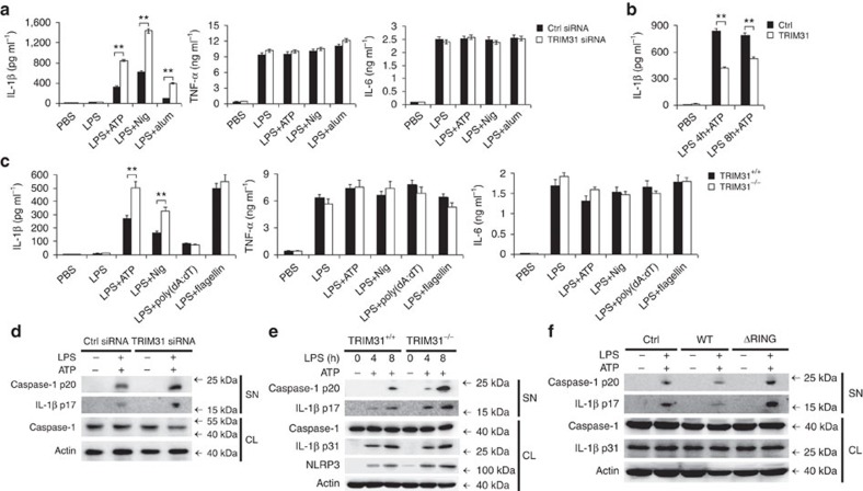
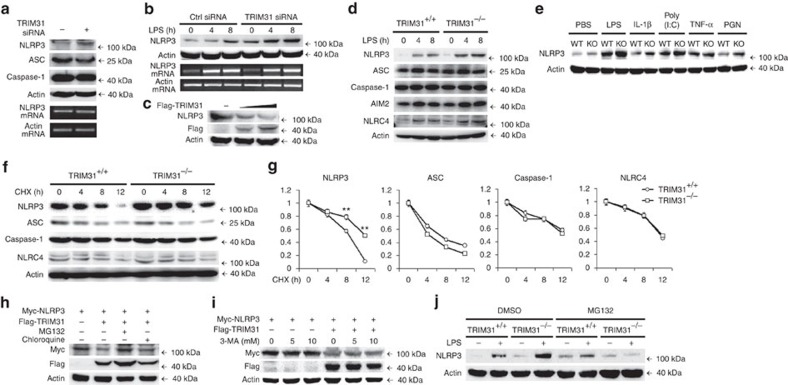
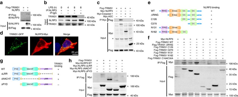
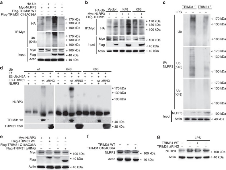
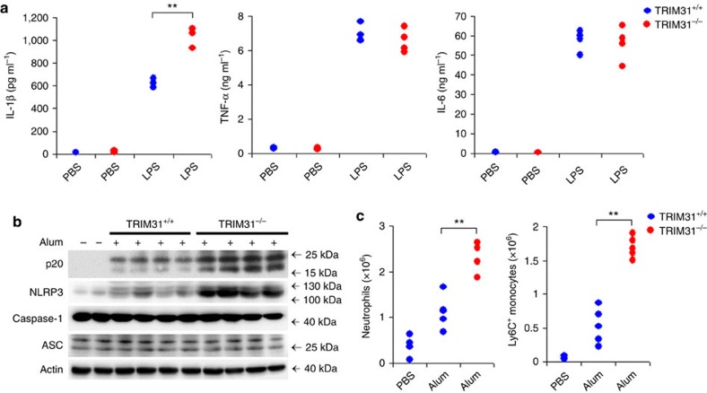
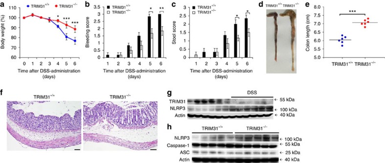
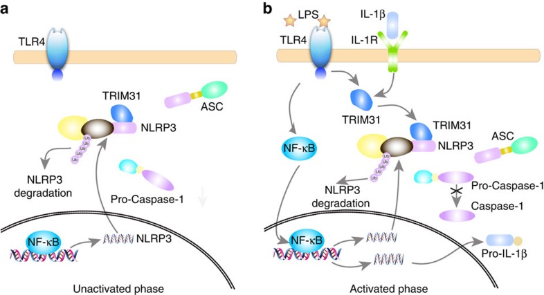
The NLRP3 inflammasome has a fundamental role in host defence against microbial pathogens and its deregulation may cause diverse inflammatory diseases. NLRP3 protein expression is a rate-limiting step for inflammasome activation, thus its expression must be tightly controlled to maintain immune homeostasis and avoid detrimental effects. However, how NLRP3 expression is regulated remains largely unknown. In this study, we identify E3 ubiquitin ligase TRIM31 as a feedback suppressor of NLRP3 inflammasome. TRIM31 directly binds to NLRP3, promotes K48-linked polyubiquitination and proteasomal degradation of NLRP3. Consequently, TRIM31 deficiency enhances NLRP3 inflammasome activation and aggravates alum-induced peritonitis in vivo. Furthermore, TRIM31 deficiency attenuates the severity of dextran sodium sulfate (DSS)-induced colitis, an inflammatory bowel diseases model in which NLRP3 possesses protective roles. Thus, our research describes a mechanism by which TRIM31 limits NLRP3 inflammasome activity under physiological conditions and suggests TRIM31 as a potential therapeutic target for the intervention of NLRP3 inflammasome related diseases.


Figures

Figure 1
Figure 1. TRIM31 specifically inhibits NLRP3 inflammasome activation.
(a) ELISA of IL-1β, TNF-α and IL-6 in supernatants from mouse peritoneal macrophages silenced of TRIM31, primed with LPS for 8 h, and followed by stimulation with ATP, Nig. or alum for 30 min. (b) ELISA of IL-1β in supernatants from THP-1 cells transfected with TRIM31 plasmid, primed with LPS for various times, and followed by stimulation with ATP for 30 min. (c) ELISA of IL-1β, TNF-α and IL-6 in supernatants of mouse peritoneal macrophages from TRIM31+/+ or TRIM31−/− mice, primed with LPS for 8 h, and followed by stimulation with ATP, Nig., poly(dA:dT) or flagellin for 30 min. (d) Immunoblot analysis of supernatants (SN) or cell lysates (CL) from mouse peritoneal macrophages silenced of TRIM31, primed with LPS, and followed by stimulation with ATP for 30 min. (e) Immunoblot analysis of supernatants (SN) or cell lysates (CL) of mouse peritoneal macrophages from TRIM31+/+ or TRIM31−/− mice, primed with LPS, and followed by stimulation with ATP for 30 min. (f) Immunoblot analysis of supernatants (SN) or cell lysates (CL) from THP-1 transfected with TRIM31 WT or ΔRING mutant, primed with LPS, and followed by stimulation with ATP for 30 min. Similar results were obtained in three independent experiments. **P<0.01. (Student's t-test). Data are representative of three experiments (mean and s.d. of six samples in ac).
Figure 2
Figure 2. TRIM31 promotes proteasomal degradation of NLRP3.
(a) Immunoblot analysis of extracts (upper panel) or RT-PCR analysis (lower panel) of mouse peritoneal macrophages silenced of TRIM31. (b) Immunoblot analysis of extracts (upper panel) or RT-PCR analysis (lower panel) of mouse peritoneal macrophages silenced of TRIM31, then stimulated for various times with LPS. (c) Immunoblot analysis of extracts from HEK293T cells transfected with increasing amount of TRIM31 expression plasmid. (d) Immunoblot analysis of extracts from TRIM31+/+ or TRIM31−/− mouse peritoneal macrophages, then stimulated for various times with LPS. (e) Immunoblot analysis of NLRP3 expression from TRIM31+/+ or TRIM31−/− mouse peritoneal macrophages, then stimulated with LPS, IL-1β, poly(I:C), TNF-α or PGN for 8 h. (f,g) Immunoblot analysis of extracts from TRIM31+/+ or TRIM31−/− mouse peritoneal macrophages stimulated with LPS for 4 h, and then treated for various times with cycloheximide (CHX). NLRP3, ASC, Caspase-1 and NLRC4 expression levels were quantitated by measuring band intensities using ‘ImageJ' software. The values were normalized to actin (g). **P<0.01. (mean and s.d. of three samples in g, Student's t-test). (h) Immunoblot analysis of extracts from HEK293T cells transfected with Myc-NLRP3 and Flag-TRIM31 expression plasmid then treated with MG132 (10 μM) or chloroquine (10 μM) for 4 h. (i) Immunoblot analysis of extracts from HEK293T cells transfected with Myc-NLRP3 and Flag-TRIM31 expression plasmid then treated with 3-MA as indicated for 4 h. (j) Immunoblot analysis of NLRP3 expression from TRIM31+/+ or TRIM31−/− mouse peritoneal macrophages stimulated with LPS for 4 h, together with DMSO or MG132 (10 μM) treatment for 4 h. Similar results were obtained in three independent experiments.
Figure 3
Figure 3. TRIM31 interacts with NLRP3.
(a) NLRP3 and Flag-tagged TRIM31 were obtained by in vitro transcription and translation. Interaction between TRIM31 and NLRP3 was assayed by mixing TRIM31 and NLRP3 together followed by IP with Flag antibody and immunoblot analysis with NLRP3 antibody. (b) Co-immunoprecipitation of endogenous TRIM31 with endogenous NLRP3 from mouse peritoneal macrophages stimulated with LPS for indicated time periods. (c) HEK293T cells expressing Flag-TRIM31 and Myc-NLRP3, Myc-Caspase-1 or Myc-ASC were lysed. Co-immunoprecipitation of Flag-TRIM31 with Myc-NLRP3 from HEK293T cells. *Non-specific band. (d) HEK293T cells transfected with GFP-TRIM31 and Myc-NLRP3 were fixed and incubated with a secondary antibody conjugated to Alexa Fluor 568. Colocalization between TRIM31 and NLRP3 was examined by Confocal microscopy. (e) Schematic diagram of TRIM31 and its truncation mutants. (f) Flag-tagged TRIM31 or its mutants and Myc-NLRP3 were individually transfected into HEK293T cells. The cell lysates were immunoprecipitated with an anti-Flag antibody and then immunoblotted with the indicated antibodies. (g) Schematic diagram of TRIM31 and its truncation mutants. (h) Myc-tagged NLRP3 or its mutants and Flag-TRIM31 were individually transfected into HEK293T cells. The cell lysates were immunoprecipitated with an anti-Flag antibody and then immunoblotted with the indicated antibodies. Similar results were obtained in three independent experiments.
Figure 4
Figure 4. TRIM31 promotes K48-linked polyubiquitination of NLRP3.
(a) Immunoblot analysis of lysates from HEK293T cells transfected with HA-tagged ubiquitin (HA-Ub), Myc-NLRP3 and TRIM31 WT or C16AC36A, followed by IP with anti-Myc, probed with anti-HA or K48-Ub. (b) Immunoblot analysis of lysates from HEK293T cells transfected with HA-tagged K48-linked ubiquitin (K48-Ub) or HA-tagged K63-linked ubiquitin (K63-Ub), Myc-NLRP3 and TRIM31, followed by IP with anti-Myc, probed with anti-HA. (c) Immunoblot analysis of lysates from TRIM31+/+ or TRIM31−/− mouse peritoneal macrophages, followed by IP with anti-NLRP3, probed with anti-Ub, K48-Ub or K63-Ub. (d) In vitro ubiquitination assay was performed in the presence of Ub (wt, K48 or K63), E1, E2-UbcH5A, NLRP3 and TRIM31 (wt or ΔRING mutant). The ubiquitination of NLRP3 was examined with NLRP3 antibody. (e) Immunoblot analysis of extracts from HEK293T cells transfected with Myc-NLRP3 and Flag-tagged TRIM31 or its mutants. (f) Immunoblot analysis of extracts from THP-1 cells transfected with TRIM31 wild type (WT) or TRIM31 C16AC36A. (g) Immunoblot analysis of extracts from THP-1 cells transfected with TRIM31 WT or TRIM31 ΔRING then stimulated with LPS for 4 h. Similar results were obtained in three independent experiments.
Figure 5
Figure 5. IL-1β and LPS induce TRIM31 expression.
(a,b) RT-PCR analysis (a) or immunoblot analysis (b) of TRIM31 expression from mouse peritoneal macrophages stimulated with LPS or IL-1β for various times. Similar results were obtained in three independent experiments. Data are representative of three experiments (mean and s.d. of six samples in a).
Figure 6
Figure 6. TRIM31 deficiency enhances IL-1β secretion and aggravates Alum-induced peritonitis in vivo.
(a) ELISA analysis of serum levels of IL-1β, TNF-α and IL-6 from TRIM31+/+ or TRIM31−/− mice after i.p. LPS injection. (b,c) TRIM31+/+ or TRIM31−/− deficiency mice were i.p. injected with alum for 12 h. PECs were lysed and analysed for the expression of Caspase-1, NLRP3 and ASC by immunoblot (b). Absolute numbers of neutrophils or Ly6C+ monocytes recruited to the peritoneum were analysed by fluorescence-activated cell sorting (five mice per group) (c). **P<0.01. (Student's t-test). Data are representative of three experiments (mean and s.d. of four to five samples in a and c).
Figure 7
Figure 7. TRIM31 deficiency ameliorates DSS-induced colitis.
Mice were given 3% DSS in their drinking water for 5 days, followed by regular drinking water. (a) Body weight, (b) stool consistency and (c) rectal bleeding score were scored daily. (dh) Mice were killed on day 6. Macroscopic appearances (d) and colon lengths (e) of the mice were measured. Histopathological changes in colon tissue were examined by H&E staining (f) Scale bars, 50μm. Immunoblot analysis of lysates from colon tissue (g,h). *P<0.05, **P<0.01, ***P<0.001 (one-way analysis of variance, ANOVA).
Figure 8
Figure 8. Working model for TRIM31 inhibiting NLRP3 inflammasome activation.
(a) In resting macrophages, the constitutively expressed TRIM31 binds to NLRP3, promotes K48-linked ubiquitination and proteasomal degradation of NLRP3, and maintains its low expression. (b) Following NLRP3 inflammasome activation, TRIM31 expression is markedly induced by LPS and IL-1β, resulting in the inhibition of NLRP3 expression and subsequent inflammasome activation.

References

    1. Schroder K. & Tschopp J. The inflammasomes. Cell 140, 821–832 (2010). - PubMed
    1. Davis B. K., Wen H. & Ting J. P. The inflammasome NLRs in immunity, inflammation, and associated diseases. Annu. Rev. Immunol. 29, 707–735 (2011). - PMC - PubMed
    1. Rathinam V. A., Vanaja S. K. & Fitzgerald K. A. Regulation of inflammasome signaling. Nat. Immunol. 13, 333–342 (2012). - PMC - PubMed
    1. Latz E., Xiao T. S. & Stutz A. Activation and regulation of the inflammasomes. Nat. Rev. Immunol. 13, 397–411 (2013). - PMC - PubMed
    1. Guo H., Callaway J. B. & Ting J. P. Inflammasomes: mechanism of action, role in disease, and therapeutics. Nat. Med. 21, 677–687 (2015). - PMC - PubMed

Publication types

MeSH terms