. 2016 Dec 8:6:38727.
doi: 10.1038/srep38727.

Peptidylarginine deiminase 1-catalyzed histone citrullination is essential for early embryo development

Affiliations

Peptidylarginine deiminase 1-catalyzed histone citrullination is essential for early embryo development

Xiaoqian Zhang et al. Sci Rep. .

Abstract

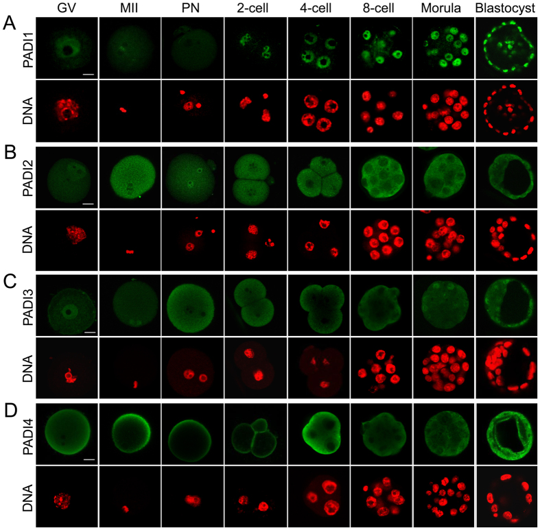
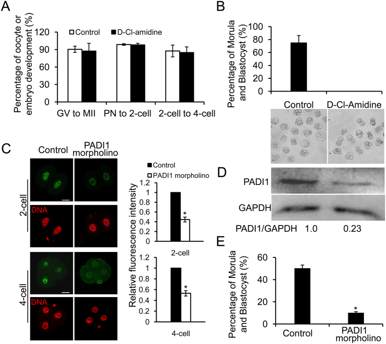
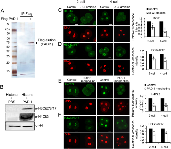
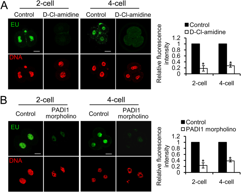
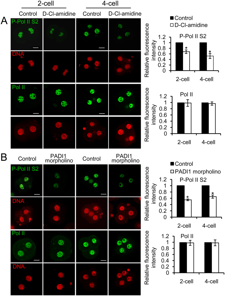
Peptidylarginine deiminase (PADI) enzymes are increasingly being associated with the regulation of chromatin structure and gene activity via histone citrullination. As one of the PADI family members, PADI1 has been mainly reported to be expressed in the epidermis and uterus, where the protein in keratinocytes is thought to promote differentiation by citrullinating filament proteins. However, the roles of PADI1 in preimplantation development have not been addressed. Using a PADI1-specific inhibitor and Padi1-morpholino knockdown, we found that citrullination of histone tails at H4R3 and H3R2/8/17 were markedly reduced in the 2- and 4-cell embryos. Consistent with this observation, early embryo development was also arrested at the 4-cell stage upon depletion of PADI1 or inhibition of PADI1 enzyme activity. Additionally, by employing 5-ethynyl uridine (EU) incorporation analysis, ablation of PADI1 function led to a dramatic decrease in overall transcriptional activity, correlating well with the reduced levels of phosphorylation of RNA Pol II at Ser2 observed at 2- or 4-cell stage of embryos under Padi1 knockdown or inhibiting PADI1. Thus, our data reveal a novel function of PADI1 during early embryo development transitions by catalyzing histone tail citrullination, which facilitates early embryo genome transactivation.


Figures

Figure 1
Figure 1. Immunofluorescence staining of PADI1, PADI2, PADI3, and PADI4.
Mouse oocytes at GV, metaphase II, and preimplantation embryos at different stages (1-cell embryos (PN), 2-cell embryos, 4-cell embryos, 8-cell embryos, morula, and blastocyst) were immunostained with antibodies specific for PADI1 (A), PADI2 (B), PADI3 (C), and PADI4 (D), respectively, and counterstained with DAPI for nuclear staining (red). Scale bar, 20 μm.
Figure 2
Figure 2. Inhibiting PADI1 activity or ablation of PADI1 blocks embryonic development in vitro beyond 4-cell stage.
(A) D-Cl-amidine treatment did not affect the rate of oocyte maturation (n = 285, the controls and n = 239, D-Cl-amidine treatment), zygote to 2-cell (n = 339, the controls and n = 342, D-Cl-amidine treatment), and 2-cell to 4-cell transitions (n = 402, the controls and n = 330, D-Cl-amidine treatment). (B) Pronuclear stage zygotes were cultured in KSOM medium supplemented without (n = 392) or with 100 μM D-Cl-amidine (n = 357) and no embryo developed to the morula or blastocyst stage. The lower panel showing the representative embryos observed by light microscopy. (C) Immunostaining confirmed that nuclear PADI1 expression significantly decreased in Padi1-MO-injected embryos at the 2- or 4-cell stage compared to the control embryos (P < 0.05). DNA was counterstained with DAPI. Scale bar, 20 μm. (D) The efficiency of Padi1-MO was verified by Western blot. Band intensity was calculated using ImageJ software, and the ratio of PADI1/GAPDH expression was normalized. (E) Depletion of PADI1 led to developmental arrest. PN zygotes were injected with Padi1-MO (n = 81) or Control-MO (n = 80), and cultured until the blastocyst stage. The embryos from each experimental group were counted and scored according to their developmental stage. Shown is the percentage of morula and blastocyst embryos from three independent experiments. *P < 0.05.
Figure 3
Figure 3. Inhibiting or knockdown PADI1 decreases histone citrullination in mouse 2-cell and 4-cell stage of embryos.
(A) Silver staining of the Flag-tagged PADI1 protein purified from Flag-PADI1 overexpressed HEK293 cells. (B) Western blotting showing the anti-H3Cit2/8/17 and anti-H4Cit3 antibodies specifically reactive with the appropriately sized band from PADI1-treated bulk histones but not from nontreated histones. Anti-histone H4 staining revealed the presence of approximately equal amounts of histone in each lane. (C–F) PN zygotes were cultured in KSOM medium for 24 h (2-cell stage) and 48 h (4-cell stage) supplemented with 100 μM D-Cl-amidine or equal volume of PBS as a control. Embryos were collected and fixed for further immunofluorescence staining with anti-H3Cit2/8/17 and anti-H4Cit3 antibodies, respectively. DNA was counterstained with DAPI. Scale bar, 20 μm. Representative images and quantification of the immunofluorescence signals from each group are shown. At least 40 embryos at each stage for each group were analyzed, and the experiments were repeated 3 times. Error bars indicate mean ± SD. *P < 0.05 vs. controls.
Figure 4
Figure 4. Immunofluorescence staining for EU incorporation.
Representative confocal images demonstrating a significant decrease of EU positive nuclear signals in the 2- or 4-cell stage of embryos treated with D-Cl-amidine (A) or Padi1-MO injection (B), compared with the control embryos, respectively. DNA was stained with Hoechst 33342 (red). Scale bar, 20 μm. Histogram on the right panel showing the quantification of EU incorporation data. Error bars indicate mean ± SD from 3 independent experiments. *P < 0.05 vs. controls.
Figure 5
Figure 5. Effect of inhibition of PADI1 or PADI1-knockdown on RNA Pol II serine 2 phosphorylation in mouse 2- and 4-cell stage embryos.
D-Cl-amidine-treated (A), and Padi1-MO injected (B) embryos (collected at the 2-cell stage and 4-cell stage separately) were immunostained with anti-RNA Pol II Ser2 (P-Pol II S2) and anti-RNA Pol II antibodies, and co-stained with DAPI for DNA. Scale bar, 20 μm. Representative images and quantification of the immunofluorescence signals from each group are shown. At least 40 embryos at each stage for each group were analyzed, and the experiments were repeated 3 times. Error bars indicate mean ± SD. *P < 0.05 vs. controls.

References

    1. Edwards R. G. Aspects of the molecular regulation of early mammalian development. Reprod Biomed Online 6, 97–113 (2003). - PubMed
    1. Hamatani T., Carter M. G., Sharov A. A. & Ko M. S. Dynamics of global gene expression changes during mouse preimplantation development. Dev Cell 6, 117–131 (2004). - PubMed
    1. Wang Q. T. et al.. A genome-wide study of gene activity reveals developmental signaling pathways in the preimplantation mouse embryo. Dev Cell 6, 133–144 (2004). - PubMed
    1. Jeong H. J. et al.. Gene expression profiling of the pre-implantation mouse embryo by microarray analysis: comparison of the two-cell stage and two-cell block. Theriogenology 66, 785–796 (2006). - PubMed
    1. Narducci M. G. et al.. TCL1 participates in early embryonic development and is overexpressed in human seminomas. Proc Natl Acad Sci USA 99, 11712–11717 (2002). - PMC - PubMed

Publication types

LinkOut - more resources