. 2017 Apr;42(5):1146-1156.
doi: 10.1038/npp.2016.275. Epub 2016 Dec 8.

A Feedforward Inhibitory Circuit Mediated by CB1-Expressing Fast-Spiking Interneurons in the Nucleus Accumbens

Affiliations

A Feedforward Inhibitory Circuit Mediated by CB1-Expressing Fast-Spiking Interneurons in the Nucleus Accumbens

William J Wright et al. Neuropsychopharmacology. 2017 Apr.

Abstract

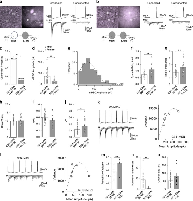
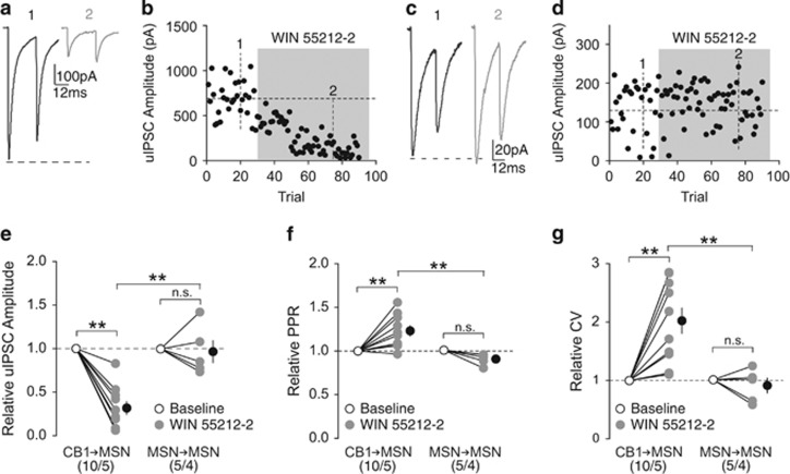
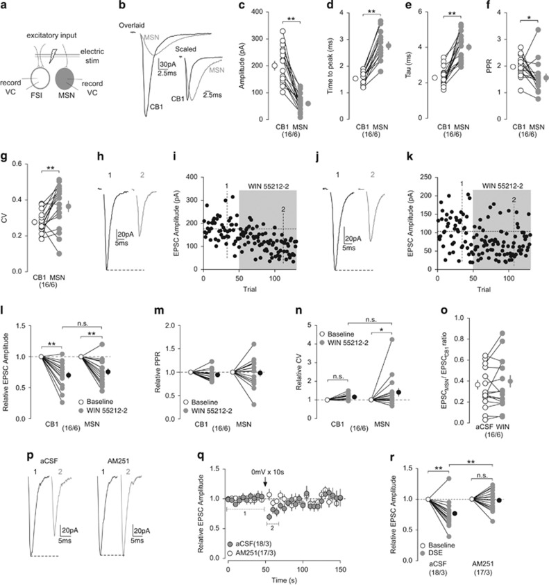
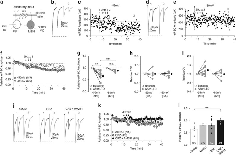
The nucleus accumbens (NAc) gates motivated behaviors through the functional output of principle medium spiny neurons (MSNs), whereas dysfunctional output of NAc MSNs contributes to a variety of psychiatric disorders. Fast-spiking interneurons (FSIs) are sparsely distributed throughout the NAc, forming local feedforward inhibitory circuits. It remains elusive how FSI-based feedforward circuits regulate the output of NAc MSNs. Here, we investigated a distinct subpopulation of NAc FSIs that express the cannabinoid receptor type-1 (CB1). Using a combination of paired electrophysiological recordings and pharmacological approaches, we characterized and compared feedforward inhibition of NAc MSNs from CB1+ FSIs and lateral inhibition from recurrent MSN collaterals. We observed that CB1+ FSIs exerted robust inhibitory control over a large percentage of nearby MSNs in contrast to local MSN collaterals that provided only sparse and weak inhibitory input to their neighboring MSNs. Furthermore, CB1+ FSI-mediated feedforward inhibition was preferentially suppressed by endocannabinoid (eCB) signaling, whereas MSN-mediated lateral inhibition was unaffected. Finally, we demonstrated that CB1+ FSI synapses onto MSNs are capable of undergoing experience-dependent long-term depression in a voltage- and eCB-dependent manner. These findings demonstrated that CB1+ FSIs are a major source of local inhibitory control of MSNs and a critical component of the feedforward inhibitory circuits regulating the output of the NAc.


Figures

Figure 1
Figure 1
Comparison of CB1-to-MSN and MSN-to-MSN inhibitory synaptic transmissions. (a) Representative images and schematic diagram (left) of paired recordings between CB1+ FSI (tdT+) and MSN (tdT), and representative uIPSC traces from functionally connected (middle) and unconnected (right) CB1-to-MSN pairs. (b) Representative images and schematic diagram (left) of paired recordings between MSN (tdT) and MSN (tdT), and representative uIPSC traces from functionally connected (middle) and unconnected (right) MSN-to-MSN pairs. (c) Summary showing CB1-to-MSN pairs exhibited a greater probability of connectivity than MSN-to-MSN pairs. (d) Summary showing amplitude of uIPSCs was greater at CB1-to-MSN synapses than MSN-to-MSN synapses. (e) Distribution of uIPSCs with a Gaussian fit showing a monomodal distribution. (f) Summary showing uIPSCs evoked at CB1-to-MSN synapses had a shorter synaptic delay than uIPSCs at MSN-to-MSN synapses. (g) Summary showing uIPSCs evoked at CB1-to-MSN synapses had shorter time to peak than uIPSCs MSN-to-MSN synapses. (h) Summary showing uIPSCs evoked at CB1-to-MSN and MSN-to-MSN synapses have similar decay kinetics. (i) Summary showing similar PPR of uIPSC responses at CB1-to-MSN and MSN-to-MSN synapses. (j) Summary showing uIPSC responses at MSN-to-MSN synapses had a greater CV than uIPSC responses at CB1-to-MSN synapses. (k) Example of uIPSC traces (left) and their hyperbolic variance mean fitted plot (right) at CB1-to-MSN synapses upon 6-pulse 20 Hz stimulation. (l) Example of uIPSC traces (left) and their hyperbolic variance mean fitted plot (right) at MSN-to-MSN synapses upon 6-pulse 20 Hz stimulation. (m) Summary showing MSN-to-MSN synapses had a higher presynaptic release probability than CB1-to-MSN synapses. (n) Summary showing CB1-to-MSN pairs had a greater number of release sites than MSN-to-MSN pairs. (o) Summary showing similar quantal size at CB1-to-MSN and MSN-to-MSN synapses. n/m represents number of cells/number of animals. *P<0.05, **p<0.01. Error bars represent SEM.
Figure 2
Figure 2
CB1 signaling preferentially suppresses inhibitory input from CB1+ FSIs. (a, b) uIPSC traces (a) and time course of uIPSC amplitude (b) from an example of functionally connected CB1-to-MSN pair before (1) and during (2) perfusion of WIN 55212-2 (5 μM). (c, d) uIPSC traces (c) and time course of uIPSC amplitude (d) from an example of functionally connected MSN-to-MSN pair before (1) and during (2) perfusion of WIN 55212-2 (5 μM). (e) Summary showing perfusion of WIN 55212-2 decreased the amplitude of uIPSCs at CB1-to-MSN synapses, whereas uIPSCs at MSN-to-MSN synapses were insensitive to WIN 55212-2. (f) Summary showing that perfusion of WIN 55212-2 increased the PPR of uIPSCs at CB1-to-MSN synapses, whereas uIPSCs at MSN-to-MSN synapses were unaffected. (g) Summary showing that perfusion of WIN 55212-2 increased the CV of uIPSC responses at CB1-to-MSN synapses, whereas uIPSCs at MSN-to-MSN synapses were unaffected. n/m represents number of cells/number of animals. **p<0.01. Error bars represent SEM.
Figure 3
Figure 3
Comparison of excitatory inputs to CB1+ FSIs and MSNs and their modulation by CB1. (a) Schematic diagram showing simultaneous dual recordings of a CB1+ FSI and an MSN upon the same electrical stimulation of presynaptic excitatory inputs. (b) Example of traces from evoked EPSCs in the simultaneously recorded CB1+ FSI (black) and MSN (gray). Overlaid traces (left) highlighting differences in amplitude and scaled traces (right) highlighting differences in activation and decay kinetics. (c) Summary showing the amplitudes of EPSCs were larger in CB1+ FSIs than MSNs. (d, e) Summary showing EPSCs evoked in CB1+ FSIs exhibited a shorter time to peak (d) and decay kinetics (e) than EPSCs in MSNs. (f) Summary showing the PPR of EPSCs evoked in CB1+ FSIs was greater than EPSCs in MSNs. (g) Summary showing the CV of EPSCs evoked in MSNs was greater than CB1+ FSIs. (h, i) EPSCs evoked in an example of CB1+ FSI (h) and the time course of the EPSC amplitudes before (1) and during (2) perfusion of WIN 55212-2 (5 μM). (j, k) EPSCs evoked in an example of MSN (j) and the time course of EPSC amplitudes (k) before (1) and during (2) perfusion of WIN 55212-2 (5 μM). Examples shown in (h–k) are from a CB1+ FSI and a MSN recorded simultaneously. (l) Summary showing that perfusion of WIN 55212-2 decreased amplitude of EPSCs evoked in CB1+ FSIs and MSNs to a similar degree. (m) Summary showing that perfusion of WIN 55212-2 did not affect the PPR of EPSCs evoked in CB1+ FSIs and MSNs. (n) Summary showing that perfusion of WIN 55212-2 increased the CV of EPSCs evoked in MSNs but not in CB1+ FSIs. (o) Summary showing that perfusion of WIN 55212-2 did not alter the EPSCMSN/EPSCCB1 ratio of simultaneously recorded pairs. (p) EPSCs evoked in an example of CB1+ FSI before (1) and after (2) a brief depolarization (0 mV for 10 s) in the absence (left) and presence (right) of AM251 (2 μM). (q) Summarized time course showing that DSE was readily induced in CB1+ FSIs and was prevented by AM251 (2 μM). (r) Summary showing that DSE decreased the amplitude of EPSCs in CB1+ FSIs that was blocked by AM251. n/m represents number of cells/number of animals. *P<0.05, **p<0.01. Error bars represent SEM.
Figure 4
Figure 4
eCB-mediated LTD of CB1-to-MSN inhibitory transmission. (a) Schematic diagram showing paired recordings of a CB1+ FSI and its connected MSN upon a LFS (2 Hz for 80 s, repeated 3 times) of synaptic inputs. (b, c) uIPSCs (b) and the time course of uIPSC amplitudes (c) from an example of CB1-to-MSN pair before (1) and following (2) LFS when MSNs were held at −55 mV during delivery of LFS. (d, e) uIPSCs (d) and the time course of uIPSC amplitudes (e) from an example of CB1-to-MSN pair before (1) and following (2) LFS when MSNs were held at −80 mV during delivery of LFS. (f) Summary showing that LFS induced LTD at CB1-to-MSN synapses when MSNs were held at −55 mV, whereas the magnitude of LTD was reduced when MSNs were held at −80 mV during LFS. (g) Summary showing that the uIPSC amplitudes at CB1-to-MSN synapses were decreased after LFS when MSNs were held at −55 mV but not −80 mV during LFS. (h) Summary showing that the PPR of uIPSCs was not significantly affected following LFS. (i) Summary showing that the CV of uIPSCs was not significantly affected following LFS. (j) Example of uIPSC traces from CB1-to-MSN pairs before (1) and following (2) LFS when MSNs were held at −55 mV during LFS delivery in the presence of AM251 (2 μM; left), CPZ (10 μM; middle), and AM251 (2 μM)+CPZ (10 μM). (k) Summary showing that LTD was abolished by AM251+CPZ, but not by AM251 or CPZ alone. (l) Summary showing that the decrease in uIPSC amplitudes following LFS (control) was partially prevented by AM251 or CPZ alone, but completely prevented by AM251+CPZ. n/m represents number of cells/number of animals. **p<0.01. Error bars represent SEM.

References

    1. Adermark L, Lovinger DM (2009). Frequency-dependent inversion of net striatal output by endocannabinoid-dependent plasticity at different synaptic inputs. J Neurosci 29: 1375–1380. - PMC - PubMed
    1. Biró AA, Holderith NB, Nusser Z (2005). Quantal size is independent of the release probability at hippocampal excitatory synapses. J Neurosci 25: 223–232. - PMC - PubMed
    1. Bolam JP, Somogyi P, Takagi H, Fodor I, Smith AD (1983). Localization of substance P-like immunoreactivity in neurons and nerve terminals in the neostriatum of the rat: a correlated light and electron microscopic study. J Neurocytol 12: 325–344. - PubMed
    1. Brog JS, Salyapongse A, Deutch AY, Zahm DS (1993). The patterns of afferent innervation of the core and shell in the “accumbens” part of the rat ventral striatum: immunohistochemical detection of retrogradely transported fluoro-gold. J Comp Neurol 338: 255–278. - PubMed
    1. Cachope R, Mateo Y, Mathur BN, Irving J, Wang H-L, Morales M et al (2012). Selective activation of cholinergic interneurons enhances accumbal phasic dopamine release: setting the tone for reward processing. Cell Rep 2: 33–41. - PMC - PubMed

Substances