Review
. 2016 Dec 6;17(12):2037.
doi: 10.3390/ijms17122037.

Integrins in the Spotlight of Cancer

Affiliations
Review

Integrins in the Spotlight of Cancer

Daniela Bianconi et al. Int J Mol Sci. .

Abstract

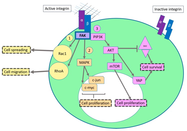
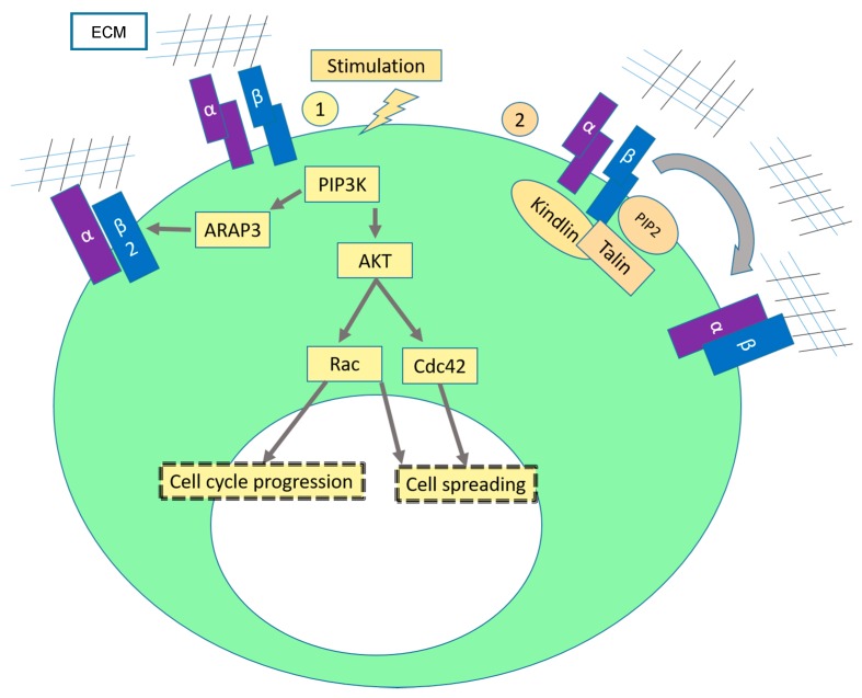
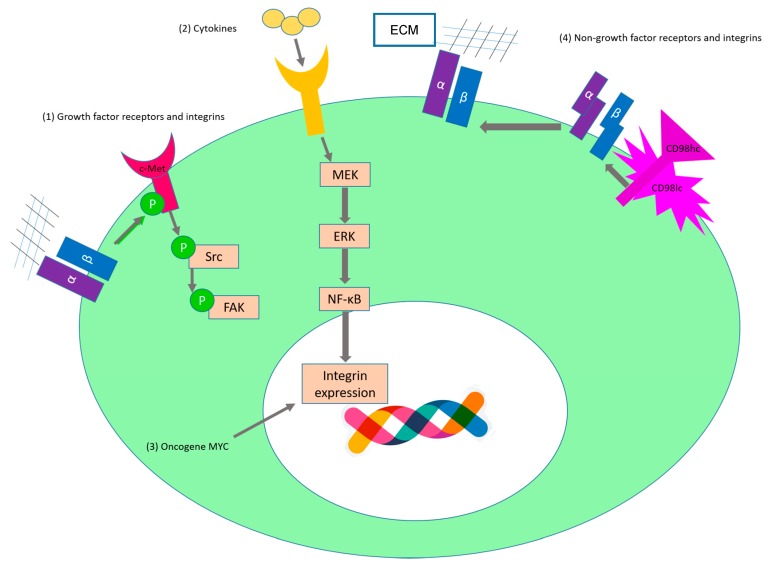
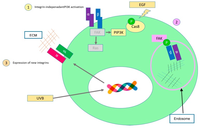
Integrins are heterodimeric cell surface receptors that bind to different extracellular ligands depending on their composition and regulate all processes which enable multicellular life. In cancer, integrins trigger and play key roles in all the features that were once described as the Hallmarks of Cancer. In this review, we will discuss the contribution of integrins to these hallmarks, including uncontrolled and limitless proliferation, invasion of tumor cells, promotion of tumor angiogenesis and evasion of apoptosis and resistance to growth suppressors, by highlighting the latest findings. Further on, given the paramount role of integrins in cancer, we will present novel strategies for integrin inhibition that are starting to emerge, promising a hopeful future regarding cancer treatment.

Keywords: angiogenesis; apoptosis; cancer; contact inhibition; integrins; invasion; locomotion; proliferation; telomerase.


Conflict of interest statement

The authors declare no conflict of interest.

Figures

Figure 1
Figure 1
Schematic representation of the integrin outside-in signaling. Through the activation of focal adhesion kinase (FAK) via integrins, Src is activated (not shown). (1) Rac1 GTPase is recruited to the plasma membrane, GDP-GTP exchange occurs and controls actin assembly in nascent protrusions [11,27]. At later stages, RhoA activity increases, leading to the formation actin stress fibers and stimulates actomyosin contractility via its downstream effector Rho-associated protein kinase (ROCK) [28]; (2) Integrin mediated FAK activation triggers the mitogen-activated protein kinase (MAPK) pathway. Different transcription factors are phosphorylated, leading to the expression of pro-proliferation genes; (3) The PIP3K/AKT pathway activation leads to enhanced translation of pro-survival and pro-proliferation genes via the mammalian target of rapamycin (mTOR) pathway. The phosphatidylinositol-3-phosphate kinase/AKR mouse thymoma kinase (PIP3K/AKT) pathway cross-talk with the Hippo pathway via Yes-associated protein (YAP). YAP is a transcription factor that can induce for example expression of the anti-apoptotic proteins survivin and Bcl-xL [29]. (Of note, there are a plethora of cross-talks between all these pathways that are not discussed here for simplication purposes). Arrows: interaction with another protein or promotion of a specific cell behavior; T-bar: inhibition; dotted boxes: effect/consequence from the signaling cascade; big ellipse in green: cell; small ellipse in white: nucleus.
Figure 2
Figure 2
Inside-out Integrin mediated cell spreading. Cell spreading is described as the deformation of the plasma membrane due to extensions of protusions, leading to the adhesion between cell and substrate [44]. (1) Upon stimulation with cytokines, PI3K is activated. On the one side, PI3K activation phorphorylates AKT and the downstream effectors Rac and Cdc42 are activated [45]. These are small GTPases that are involved in the reorganization of the cytoskeleton which leads to the formation of lamelipodia and filipodia. On the other side, it was shown that PI3K induces activity of the β2 integrin subunit via ARAP3 (Arf GAP and Rho GAP with ankyrin repeat and PH domain 3) [46]. ARAP3 is Rap-regulated GTPase-activating protein for RhoA and Arf6 which are resposible for actin modulation [47,48,49]; (2) Accumulation of PIP2 leads to the recruitment of talin to the plasma membrane, enabling a physical interaction between talin and the β1 and β3 integrin subunits which induces a conformational change in the integrin heterodimers that increases integrin affinity to its ligand. Kindlins are also essential for the inside-out activation of integrins [50]. In fibroblasts, kindlin-2 binds and activates FAK, inducing formation of lamellipodia [51]. Arrows: interaction with another protein or promotion of a specific cell behavior; T-bar: inhibition; dotted boxes: effect/consequence from the signaling cascade; big ellipse in green: cell; small ellipse in white: nucleus.
Figure 3
Figure 3
Role of integrins in tumor cell proliferation. (1) The crosstalk between integrins and growth factor receptors (summarized in Table 1) promotes cancer cell proliferation. For instance, in ovarian cancer cells c-Met is activated via phosphorylation upon binding of α5β1 to fibronectin [65]. This activation leads to activation of Src and FAK and promotes proliferation and invasion [65]; (2) Tumor necrosis factor α (TNF-α) leads to an increased expression of integrins such as α2β1 and αvβ3 [93,101]; (3) The oncogene MYC (v-myc avian myelocytomatosis viral oncogene homolog) can bind to promoter elements of α1β1 and leads to enhanced transcription of this integrin [87]; (4) Integrins can interact with transmembrane receptors such as CD98hc. CD98hc modulates integrin signaling by o conferring integrins with “stiffness” that allows them to trigger their signaling pathway [75]. (Of note, the DNA cartoon was designed by Freepik). Big ellipse in green: cell; small ellipse in white: nucleus.
Figure 4
Figure 4
Schematic representation of some integrin-mediated mechanisms to avoid anoikis. (1) Growth factors can phosphorylate caspase-8 and this allows interaction and activation with PI3K; (2) Integrins are endocytosed and integrin signaling continues in the endosomes via interaction with FAK; (3) Integrins that are not expressed in a specific tissue are upregulated. These integrins can bind to new components of the extracellular matrix (ECM) and start signaling. (Of note, the DNA cartoon was designed by Freepik). Big ellipse in green: cell; small ellipse in white: nucleus.

References

    1. Hynes R.O. Integrins: Bidirectional, allosteric signaling machines. Cell. 2002;110:673–687. doi: 10.1016/S0092-8674(02)00971-6. - DOI - PubMed
    1. Ginsberg M.H. Integrin activation. BMB Rep. 2014;47:655–659. doi: 10.5483/BMBRep.2014.47.12.241. - DOI - PMC - PubMed
    1. Sebe-Pedros A., Roger A.J., Lang F.B., King N., Ruiz-Trillo I. Ancient origin of the integrin-mediated adhesion and signaling machinery. Proc. Natl. Acad. Sci. USA. 2010;107:10142–10147. doi: 10.1073/pnas.1002257107. - DOI - PMC - PubMed
    1. Johnson M.S., Lu N., Denessiouk K., Heino J., Gullberg D. Integrins during evolution: Evolutionary trees and model organisms. Biochim. Biophys. Acta. 2009;1788:779–789. doi: 10.1016/j.bbamem.2008.12.013. - DOI - PubMed
    1. Hanahan D., Weinberg R.A. The hallmarks of cancer. Cell. 2000;100:57–70. doi: 10.1016/S0092-8674(00)81683-9. - DOI - PubMed

MeSH terms