Review
. 2016 Dec 6;17(12):2045.
doi: 10.3390/ijms17122045.

Cell Death in Chondrocytes, Osteoblasts, and Osteocytes

Affiliations
Review

Cell Death in Chondrocytes, Osteoblasts, and Osteocytes

Toshihisa Komori. Int J Mol Sci. .

Abstract

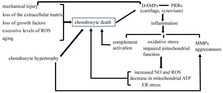
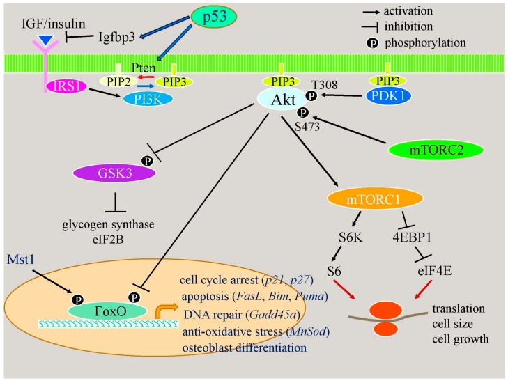
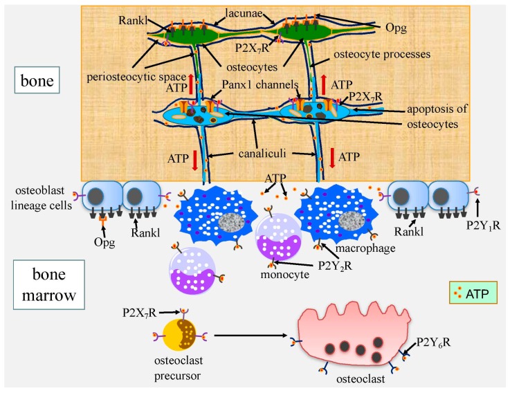
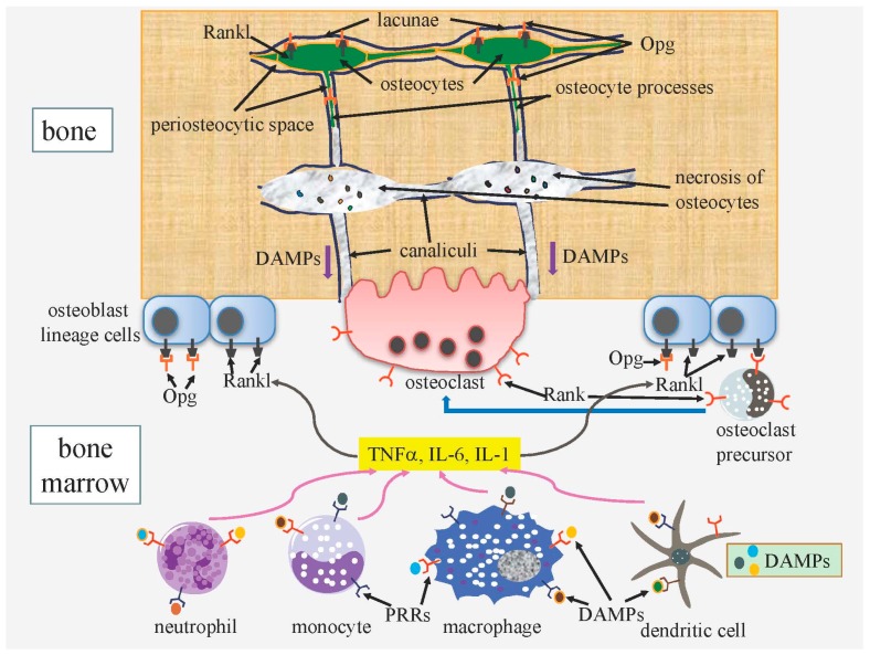
Cell death in skeletal component cells, including chondrocytes, osteoblasts, and osteocytes, plays roles in skeletal development, maintenance, and repair as well as in the pathogenesis of osteoarthritis and osteoporosis. Chondrocyte proliferation, differentiation, and apoptosis are important steps for endochondral ossification. Although the inactivation of P53 and RB is involved in the pathogenesis of osteosarcomas, the deletion of p53 and inactivation of Rb are insufficient to enhance chondrocyte proliferation, indicating the presence of multiple inhibitory mechanisms against sarcomagenesis in chondrocytes. The inflammatory processes induced by mechanical injury and chondrocyte death through the release of danger-associated molecular patterns (DAMPs) are involved in the pathogenesis of posttraumatic osteoarthritis. The overexpression of BCLXL increases bone volume with a normal structure and maintains bone during aging by inhibiting osteoblast apoptosis. p53 inhibits osteoblast proliferation and enhances osteoblast apoptosis, thereby reducing bone formation, but also exerts positive effects on osteoblast differentiation through the Akt-FoxOs pathway. Apoptotic osteocytes release ATP, which induces the receptor activator of nuclear factor κ-B ligand (Rankl) expression and osteoclastogenesis, from pannexin 1 channels. Osteocyte death ultimately results in necrosis; DAMPs are released to the bone surface and promote the production of proinflammatory cytokines, which induce Rankl expression, and osteoclastogenesis is further enhanced.

Keywords: ATP; BCLXL; DAMPs; FoxO; Rankl; Rb; apoptosis; necrosis; osteoarthritis; p53.


Conflict of interest statement

The author declares no conflict of interest.

Figures

Figure 1
Figure 1
Chondrocyte death in osteoarthritis. Mechanical injury, the loss of the extracellular matrix, loss of growth factors, and excessive levels of reactive oxygen species (ROS) induce chondrocyte death in articular cartilage. Chondrocyte hypertrophy in articular cartilage induces the destruction of the cartilage matrix through the induction of matrix metalloproteinases (MMPs) and aggrecanases, and leads to chondrocyte apoptosis. The number of dead chondrocytes also increases in articular cartilage during aging. DAMPs (danger-associated molecular patterns) released by mechanical injury and chondrocyte secondary necrosis initiate non-infectious inflammatory responses through PRRs (pattern recognition receptors) expressed in osteoarthritis (OA) cartilage and the synovium, causing complement activation, oxidative stress, impaired mitochondrial function, and the induction of MMPs and aggrecanases. Complement activation, an increase in nitric oxide (NO) and ROS, a decrease in mitochondrial ATP, and endoplasmic reticulum (ER) stress further enhance chondrocyte death, resulting in the progression of osteoarthritis. The contribution of chondrocyte death to inflammatory processes needs to be evaluated. The inflammatory processes induced by mechanical injury contribute to the development of posttraumatic OA; however, their significance in non-posttraumatic OA remains unclear.
Figure 2
Figure 2
Acceleration of osteoblast differentiation through the p53–Akt–FoxO pathway. p53 induces the expression of Igfbp3 and Pten. Igfbp3 inhibits IGF/insulin binding to the receptor, and Pten dephosphorylates PIP3 to PIP2. PIP3 recruits Akt to the cell membrane and Akt is activated by PDK1 and mTORC2. The decrease in PIP3 inactivates Akt. Akt regulates metabolism, cell growth, cell survival, cell proliferation, and cell differentiation through GSK3, mTORC1, and FoxOs. Akt phosphorylates FoxOs to inactivate them. FoxOs are involved in cell cycle arrest, apoptosis, DNA repair, anti-oxidative stress, and osteoblast differentiation.
Figure 3
Figure 3
Induction of bone resorption by ATP released from apoptotic osteocytes. In apoptotic osteocytes, active Panx1 channels, which release ATP, are formed by the caspase-mediated cleavage of the C terminus. P2X7 receptor (P2X7R), which is activated by extracellular ATP, forms a complex with Panx1 and enhances ATP release. Extracellular ATP recruits macrophages and monocytes through P2Y2R, increases nuclear factor κ-B ligand (Rankl) expression in osteocytes through P2X7R and in osteoblasts through P2Y1R, increases osteoclast survival through P2Y6R, and enhances the membrane fusion of osteoclast precursor cells to form multinucleated osteoclasts through P2X7R. The up-regulation of Rankl in osteocytes will reduce the release of osteoprotegerin (Opg), which is secreted by osteocytes, to the bone surface. It has been shown that Panx1 and P2X7R are required for Rankl up-regulation in osteocytes in fatigued bone. However, the functions of ATP released from apoptotic osteocytes shown here have not yet been directly proven in damaged bone.
Figure 4
Figure 4
A proposed scheme for the enhanced osteoclastogenesis by osteocyte death. Any type of osteocyte death ultimately results in necrosis because dead osteocytes are not phagocytosed. DAMPs (danger-associated molecular patterns) released from necrotic osteocytes pass through canaliculi and reach the bone surface. Macrophages, dendritic cells, neutrophils, and monocytes are stimulated by DAMPs through PRRs (pattern recognition receptors), and produce TNFα, IL-6, and IL-1, which stimulate the expression of Rankl in osteoblast lineage cells. Osteoclast precursors differentiate into osteoclasts through Rankl-Rank signaling. Opg released from osteocytes to the bone surface negatively regulates osteoclastogenesis and osteoclast activity. This scheme is predicted from previous studies, but has not been directly proven in damaged bone.

References

    1. Yoshida C.A., Yamamoto H., Fujita T., Furuichi T., Ito K., Inoue K., Yamana K., Zanma A., Takada K., Ito Y., et al. Runx2 and Runx3 are essential for chondrocyte maturation, and Runx2 regulates limb growth through induction of Indian hedgehog. Genes Dev. 2004;18:952–963. doi: 10.1101/gad.1174704. - DOI - PMC - PubMed
    1. Inada M., Yasui T., Nomura S., Miyake S., Deguchi K., Himeno M., Sato M., Yamagiwa H., Kimura T., Yasui N., et al. Maturational disturbance of chondrocytes in Cbfa1-deficient mice. Dev. Dyn. 1999;214:279–290. doi: 10.1002/(SICI)1097-0177(199904)214:4<279::AID-AJA1>3.0.CO;2-W. - DOI - PubMed
    1. St-Jacques B., Hammerschmidt M., McMahon A.P. Indian hedgehog signaling regulates proliferation and differentiation of chondrocytes and is essential for bone formation. Genes Dev. 1999;13:2072–2086. doi: 10.1101/gad.13.16.2072. - DOI - PMC - PubMed
    1. Iwamoto M., Kitagaki J., Tamamura Y., Gentili C., Koyama E., Enomoto H., Komori T., Pacifici M., Enomoto-Iwamoto M. Runx2 expression and action in chondrocytes are regulated by retinoid signaling and parathyroid hormone-related peptide (PTHrP) Osteoarthr. Cartil. 2003;11:6–15. doi: 10.1053/joca.2002.0860. - DOI - PubMed
    1. Li T.F., Dong Y., Ionescu A.M., Rosier R.N., Zuscik M.J., Schwarz E.M., O’Keefe R.J., Drissi H. Parathyroid hormone-related peptide (PTHrP) inhibits Runx2 expression through the PKA signaling pathway. Exp. Cell Res. 2004;299:128–136. doi: 10.1016/j.yexcr.2004.05.025. - DOI - PubMed