

Comparison of liver volumetry on contrast-enhanced CT images: one semiautomatic and two automatic approaches
- PMID: 27929487
- PMCID: PMC5690519
- DOI: 10.1120/jacmp.v17i6.6485
Comparison of liver volumetry on contrast-enhanced CT images: one semiautomatic and two automatic approaches
Abstract
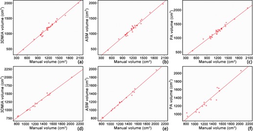
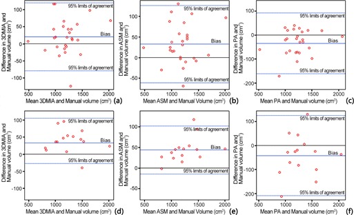
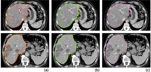
This study was to evaluate the accuracy, consistency, and efficiency of three liver volumetry methods- one interactive method, an in-house-developed 3D medical Image Analysis (3DMIA) system, one automatic active shape model (ASM)-based segmentation, and one automatic probabilistic atlas (PA)-guided segmentation method on clinical contrast-enhanced CT images. Forty-two datasets, including 27 normal liver and 15 space-occupying liver lesion patients, were retrospectively included in this study. The three methods - one semiautomatic 3DMIA, one automatic ASM-based, and one automatic PA-based liver volumetry - achieved an accuracy with VD (volume difference) of -1.69%, -2.75%, and 3.06% in the normal group, respectively, and with VD of -3.20%, -3.35%, and 4.14% in the space-occupying lesion group, respectively. However, the three methods achieved an efficiency of 27.63 mins, 1.26 mins, 1.18 mins on average, respectively, compared with the manual volumetry, which took 43.98 mins. The high intraclass correlation coefficient between the three methods and the manual method indicated an excel-lent agreement on liver volumetry. Significant differences in segmentation time were observed between the three methods (3DMIA, ASM, and PA) and the manual volumetry (p < 0.001), as well as between the automatic volumetries (ASM and PA) and the semiautomatic volumetry (3DMIA) (p < 0.001). The semiautomatic interactive 3DMIA, automatic ASM-based, and automatic PA-based liver volum-etry agreed well with manual gold standard in both the normal liver group and the space-occupying lesion group. The ASM- and PA-based automatic segmentation have better efficiency in clinical use.
© 2016 The Authors.
Figures
References
-
- International agency for research on cancer . Globocan 2012: Estimated Cancer Incidence, Mortality and Prevalence Worldwide in 2012. All Cancers (excluding non‐melanoma skin cancer) Estimated Incidence, Mortality and Prevalence Worldwide in 2012. Available from: http://globocan.iarc.fr/Pages/fact_sheets_cancer.aspx. Accessed 12 May, 2016.
-
- Lau WY and Lai EC. Hepatocellular carcinoma: current management and recent advances. Hepatob Pancreat Dis. 2008;7(3):237–57. - PubMed
-
- El‐Serag HB. Current concepts: hepatocellular carcinoma. New Engl J Med. 2011;365(12):1118–27. - PubMed
-
- Frericks BB, Caldarone FC, Nashan B, et al. 3D CT modeling of hepatic vessel architecture and volume calculation in living donated liver transplantation. Eur Radiol. 2004;14(2):326–33. - PubMed
MeSH terms
LinkOut - more resources
Full Text Sources
Other Literature Sources
Medical
