. 2017 Jan 17;16(2):179-188.
doi: 10.1080/15384101.2016.1261226. Epub 2016 Dec 8.

Exogenous expression of SAMHD1 inhibits proliferation and induces apoptosis in cutaneous T-cell lymphoma-derived HuT78 cells

Affiliations

Exogenous expression of SAMHD1 inhibits proliferation and induces apoptosis in cutaneous T-cell lymphoma-derived HuT78 cells

Karthik M Kodigepalli et al. Cell Cycle. .

Abstract

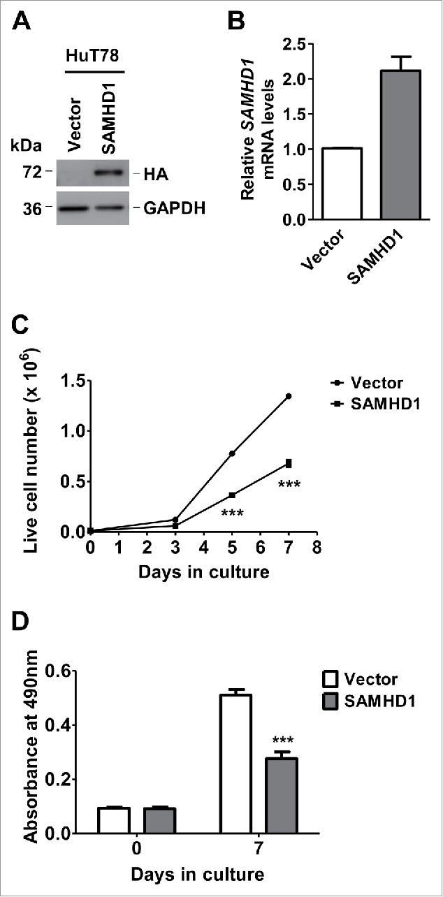
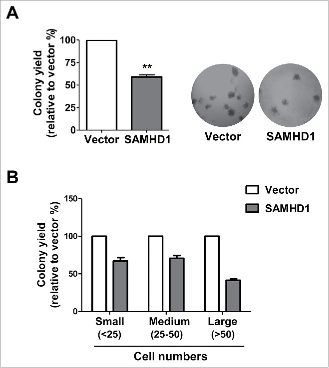
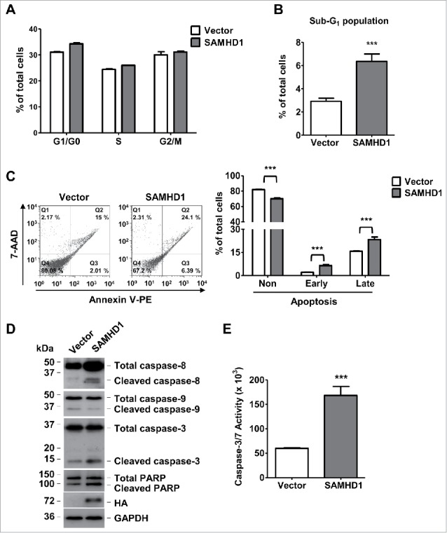
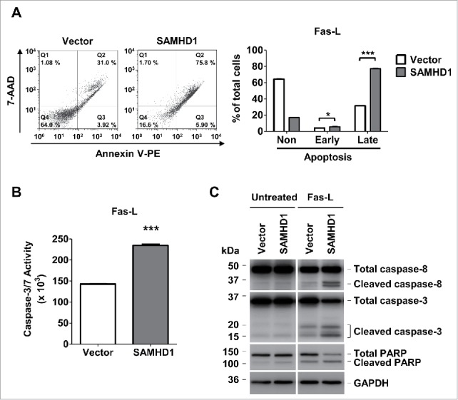
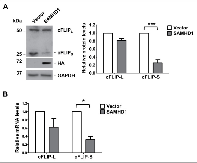
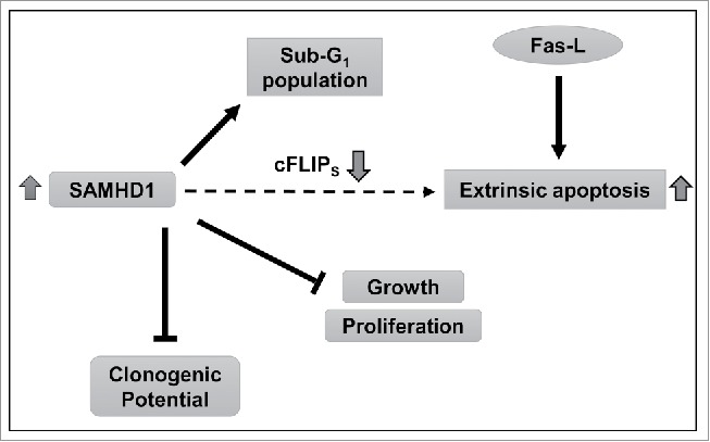
Sterile α motif and HD domain-containing protein 1 (SAMHD1) is a mammalian dNTP hydrolase (dNTPase) that regulates intracellular dNTP balance. We have previously reported that SAMHD1 mRNA and protein levels are significantly downregulated in CD4+ T-cells of patients with cutaneous T-cell lymphoma (CTCL), a disease characterized by infiltration of neoplastic CD4+ T-lymphocytes into the skin. However, functional significance of SAMHD1 in CTCL development and progression remains unknown. Here we investigate the mechanism by which SAMHD1 induces apoptosis in CTCL-derived CD4+ T-cells. We stably expressed exogenous SAMHD1 in the CTCL-derived HuT78 T-cell line containing a very low level of endogenous SAMHD1 protein. We found that low-level exogenous expression of SAMHD1 led to a significant reduction in HuT78 cell growth, proliferation, and colony formation. Exogenous SAMHD1 expression in HuT78 cells also resulted in increased spontaneous and Fas ligand (Fas-L)-induced apoptosis levels via activation of the extrinsic pathway, including caspase-8, -3 and -7. Additionally, increased SAMHD1 significantly reduced the protein and mRNA expression of the short isoform of cFLIP (cFLIPS), an important negative regulator of Fas-L-mediated apoptotic signaling. Our results indicate that exogenous SAMHD1 expression inhibits HuT78 cell growth and proliferation in part by increasing apoptosis. These findings implicate that SAMHD1 acts as an inhibitor in CTCL cell growth, suggesting that downregulation of SAMHD1 expression in neoplastic T-cells can facilitate uncontrolled cell proliferation.

Keywords: Fas-L; SAMHD1; apoptosis; cFLIP; cell proliferation; lymphoma.


Figures

Figure 1.
Figure 1.
Exogenous SAMHD1 expression inhibits HuT78 cell growth and proliferation. HuT78 cells expressing hemagglutinin (HA)-tagged SAMHD1 were generated via lentiviral vector-mediated transduction. (A) Exogenous expression of SAMHD1 was validated by immunoblotting analysis using antibodies to HA (SAMHD1) and GAPDH (loading control). (B) Total RNA was isolated from HuT78 vector or SAMHD1-expressing cells and qPCR analysis was performed to quantify the relative SAMHD1 mRNA levels. GAPDH mRNA levels were quantified as internal control. (C) HuT78 vector control and SAMHD1 expressing cells were seeded in triplicate in 24-well plates at a density of 1 × 104 cells/well in 1 ml culture media on day 0 and live cells were counted on indicated days via trypan-blue exclusion method. (D) Cell lines were seeded on day 0 in 4 replicates in 96-well plates at density of 1 × 103 cells/well in 100 µl and cell proliferation was determined on indicated days utilizing an MTS-based proliferation assay. All the data presented are representative of 3 or more independent experiments. (C-D) ***, p < 0.0001.
Figure 2.
Figure 2.
Exogenous SAMHD1 expression in HuT78 cells results in reduced colony formation. HuT78 vector control, and SAMHD1-expressing cells were plated in duplicate in 6-well plates at a density of 1 × 103 cells/well in 1 ml methyl cellulose medium and were left in culture for 10 d. (A) Numbers of colonies were counted after 10 d following standard counting criteria (all colonies with >25 cells were counted). Data is presented as % of colony yield relative to vector control cells (left panel). **, p < 0.005, Representative images (20 × magnification) of colonies formed after 10 d (right panel). (B) Numbers of colonies of different sizes were counted after 10 d based on following standard counting criteria (small: <25 cells; medium: 25–50 cells, and large: >50 cells). Data is presented as percentages of colony yield relative to vector control cells. All the data presented are representative of 3 independent experiments.
Figure 3.
Figure 3.
Exogenous SAMHD1 expression induces apoptosis and caspase-3/7 activity via activation of extrinsic apoptosis signaling. HuT78 vector control and SAMHD1 expressing cells were seeded at density of 2 × 104 cells per 6-cm dish containing 5 ml growth media on day 0 following trypan-blue exclusion method. (A) Seven days post-seeding, cells in triplicate, were stained with propidium iodide (PI) and cell cycle analysis was performed via flow cytometry. Percentages of cells in G1/G0, S, and G2/M phases of cell cycle are presented. (B) Percentage of cells in Sub-G1 phase are presented. (C) HuT78 vector control and SAMHD1-expressing cells were seeded at density of 2 × 104 cells per 6-cm dish containing 5 ml growth media on day 0 following trypan-blue exclusion method. Seven days post-seeding, cells were stained with annexin V-PE and 7-AAD in triplicate and analyzed by flow cytometry. Representative flow cytometric profiles are presented (left panel). The percentages of non-apoptotic cells, early apoptotic cells, and late apoptotic cells were quantified as presented (right panel). (D) On day 7 post-seeding, cell lysates from all cell lines were collected and western analysis was performed using the antibodies to caspase-8, caspase-9, caspase-3, PARP, HA (SAMHD1), and GAPDH (loading control). (E) On day 7 post-seeding, 1 × 104 cells per cell line were collected in 4 replicates and incubated in 100 µl of Caspase-Glo 3/7 reagent at room temperature in dark for 1 hour. Caspase-3/7 activity was then determined by measuring luminescence values. All the data presented are representative of 3 independent experiments. B, C, E, ***, p < 0.001.
Figure 4.
Figure 4.
Exogenous SAMHD1 expression in HuT78 cells significantly increases Fas-L induced apoptosis and caspase-3/7 activity. (A) HuT78 vector control and SAMHD1-expressing cells were treated with 100 ng/ml Fas-L for 48 hours. After 48 hours of treatment, all the cells in triplicate were stained with annexin V-PE and 7-AminoactinomycinD (7-AAD) followed by flow cytometry. Representative flow cytometric profiles are presented (left panel). The percentage of non-apoptotic cells, early apoptotic cells, and late apoptotic cells were quantified as presented (right panel). (B) HuT78 vector control and SAMHD1-expressing cells were treated with 100 ng/ml Fas-L for 48 hours. Post-treatment, 1 × 104 cells per cell line were collected in triplicate and incubated in 100 µl of Caspase-Glo 3/7 reagent at room temperature in dark for 1 hour. Caspase-3/7 activity was then determined by measuring luminescence values. C, HuT78 vector control and SAMHD1-expressing cells were treated with Fas-L (100 ng/ml) for 48 hours. Post-treatment, cell lysates from all cell lines were collected and immunoblotting was performed using the antibodies to caspase-8, caspase-3, PARP, and GAPDH (loading control). All the data presented are representative of 3 independent experiments. A–B, ***, p < 0.001.
Figure 5.
Figure 5.
Exogenous SAMHD1 expression in HuT78 cells reduces the levels of cFLIPS protein and mRNA. HuT78 vector control and SAMHD1-expressing cells were seeded at density of 2 × 104 cells per 6-cm dish containing 5 ml growth media on day 0 following trypan-blue exclusion method. (A) On day 7 post-seeding, cell lysates were collected and immunoblotting analysis was performed for cFLIP, HA (SAMHD1) and GAPDH (loading control) (left panel). Densitometry was performed using ImageJ to quantify the relative cFLIPL and cFLIPS protein levels (right panel). ***, p < 0.001 (B) On day 7 post-seeding, total RNA was isolated from HuT78 vector or SAMHD1-expressing cells and qPCR analysis was performed to quantify the relative cFLIPL and cFLIPS mRNA levels. GAPDH mRNA levels were quantified as internal control. All the data presented are representative of 3 independent experiments. *, p < 0.05.
Figure 6.
Figure 6.
Summary of the findings and proposed mechanisms. Our data indicate that increased SAMHD1 expression inhibits growth and proliferation in CTCL-derived CD4+ T-cells by inducing spontaneous and Fas-L stimulated apoptosis. SAMHD1 expression also diminishes colony forming potential in these cells. Furthermore, increased SAMHD1 expression also significantly reduces the mRNA and protein levels of cFLIPS, a key anti-apoptotic protein. Complete mechanisms by which SAMHD1 regulates cFLIPS expression, apoptosis and colony formation in HuT78 cells remain to be understood.

References

    1. Goldstone DC, Ennis-Adeniran V, Hedden JJ, Groom HC, Rice GI, Christodoulou E, Walker PA, Kelly G, Haire LF, Yap MW, et al.. HIV-1 restriction factor SAMHD1 is a deoxynucleoside triphosphate triphosphohydrolase. Nature 2011; 480:379-82; PMID:22056990; http://dx.doi.org/ 10.1038/nature10623 - DOI - PubMed
    1. Hrecka K, Hao C, Gierszewska M, Swanson SK, Kesik-Brodacka M, Srivastava S, Florens L, Washburn MP, Skowronski J. Vpx relieves inhibition of HIV-1 infection of macrophages mediated by the SAMHD1 protein. Nature 2011; 474:658-61; PMID:21720370; http://dx.doi.org/ 10.1038/nature10195 - DOI - PMC - PubMed
    1. Laguette N, Sobhian B, Casartelli N, Ringeard M, Chable-Bessia C, Ségéral E, Yatim A, Emiliani S, Schwartz O, Benkirane M. SAMHD1 is the dendritic- and myeloid-cell-specific HIV-1 restriction factor counteracted by Vpx. Nature 2011; 474:654-7; PMID:21613998; http://dx.doi.org/ 10.1038/nature10117 - DOI - PMC - PubMed
    1. Powell RD, Holland PJ, Hollis T, Perrino FW. Aicardi-Goutieres syndrome gene and HIV-1 restriction factor SAMHD1 is a dGTP-regulated deoxynucleotide triphosphohydrolase. J Biol Chem 2011; 286:43596-600; PMID:22069334; http://dx.doi.org/ 10.1074/jbc.C111.317628 - DOI - PMC - PubMed
    1. Franzolin E, Pontarin G, Rampazzo C, Miazzi C, Ferraro P, Palumbo E, Reichard P, Bianchi V. The deoxynucleotide triphosphohydrolase SAMHD1 is a major regulator of DNA precursor pools in mammalian cells. Proc Natl Acad Sci U S A 2013; 110:14272-7; PMID:23858451; http://dx.doi.org/ 10.1073/pnas.1312033110 - DOI - PMC - PubMed

MeSH terms

Substances

LinkOut - more resources