. 2017 Feb;174(4):287-301.
doi: 10.1111/bph.13688. Epub 2017 Jan 24.

AP39, a mitochondria-targeting hydrogen sulfide (H2 S) donor, protects against myocardial reperfusion injury independently of salvage kinase signalling

Affiliations

AP39, a mitochondria-targeting hydrogen sulfide (H2 S) donor, protects against myocardial reperfusion injury independently of salvage kinase signalling

Qutuba G Karwi et al. Br J Pharmacol. 2017 Feb.

Abstract

Background and purpose: H2 S protects myocardium against ischaemia/reperfusion injury. This protection may involve the cytosolic reperfusion injury salvage kinase (RISK) pathway, but direct effects on mitochondrial function are possible. Here, we investigated the potential cardioprotective effect of a mitochondria-specific H2 S donor, AP39, at reperfusion against ischaemia/reperfusion injury.

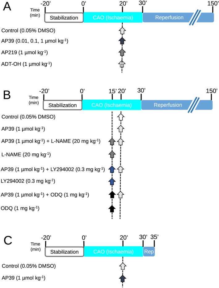
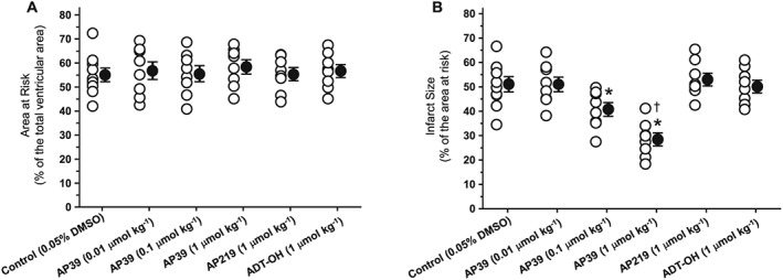
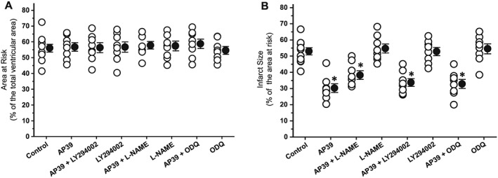
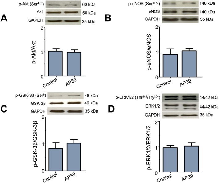
Experimental approach: Anaesthetized rats underwent myocardial ischaemia (30 min)/reperfusion (120 min) with randomization to receive interventions before reperfusion: vehicle, AP39 (0.01, 0.1, 1 μmol·kg-1 ), or control compounds AP219 and ADT-OH (1 μmol·kg-1 ). LY294002, L-NAME or ODQ were used to investigate the involvement of the RISK pathway. Myocardial samples harvested 5 min after reperfusion were analysed for RISK protein phosphorylation and isolated cardiac mitochondria were used to examine the direct mitochondrial effects of AP39.

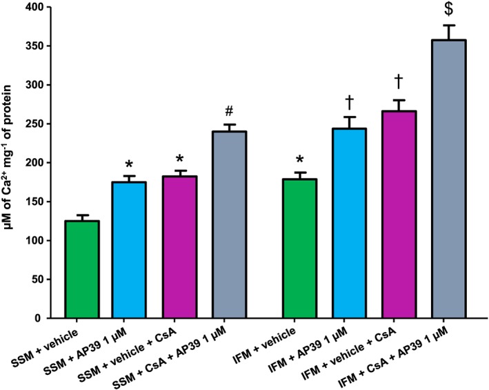
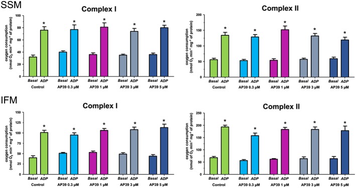
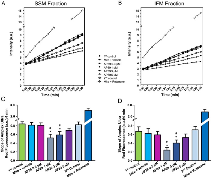
Key results: AP39, dose-dependently, reduced infarct size. Inhibition of either PI3K/Akt, eNOS or sGC did not affect this effect of AP39. Western blot analysis confirmed that AP39 did not induce phosphorylation of Akt, eNOS, GSK-3β or ERK1/2. In isolated subsarcolemmal and interfibrillar mitochondria, AP39 significantly attenuated mitochondrial ROS generation without affecting respiratory complexes I or II. Furthermore, AP39 inhibited mitochondrial permeability transition pore (PTP) opening and co-incubation of mitochondria with AP39 and cyclosporine A induced an additive inhibitory effect on the PTP.

Conclusion and implications: AP39 protects against reperfusion injury independently of the cytosolic RISK pathway. This cardioprotective effect could be mediated by inhibiting PTP via a cyclophilin D-independent mechanism. Thus, selective delivery of H2 S to mitochondria may be therapeutically applicable for employing the cardioprotective utility of H2 S.


Figures

Figure 1
Figure 1
Experimental protocols: Animals underwent 30 min of ischaemia followed by 2 h of reperfusion. Infarct size was determined using Evans' blue/TTC staining technique. Infarction was reported as a percentage of the AAR (I/AAR %). (A) AP39 dose effect on infarct size: Animals were randomly assigned to be treated with either vehicle or AP39 or the controls (AP219 or ADT‐OH) at 10 min before reperfusion. (B) Mechanistic study: Rats were randomised to receive the pharmacological inhibitors, namely LY294002, L‐NAME and ODQ, at 15 min before reperfusion with or without AP39 applied at 10 min before reperfusion. Control group only received the vehicle (0.05% DMSO) 10 min before reperfusion. (C) Myocardium sampling protocol: Rats were randomised to receive either vehicle (0.05% DMSO) or AP39 10 minutes before reperfusion. Myocardial biopsies were harvested at 5 minutes of reperfusion from the left ventricle. Arrows indicate the time of the pharmacological interventions.
Figure 2
Figure 2
Infarct‐limiting effect of AP39 at reperfusion: (A) area at risk (AAR) reported as a percentage of the total ventricular volume. (B) Infarct size presented as a percentage of the AAR. Data were analysed using one‐way ANOVA with Neuman Keuls post hoc test and presented as mean ± SEM, n = 8 for all groups except the control group (0.05% DMSO) where n = 10. The mean of the infarct size for each group is represented by a filled circle (with error bars) next to the individual values (open circles). * P < 0.05 versus control, P < 0.05 versus AP39 0.1 μmol·kg−1.
Figure 3
Figure 3
Effect of pharmacological inhibitors of the RISK pathway on infarct‐limitation by AP39: (A) risk zone measurements of experimental groups expressed as a percentage of the total ventricular area. (B) Myocardial infarction data are expressed as a percentage of the risk zone. Individual animal data in each group are represented by empty circles while the mean of infarct size is presented by a full circle. Data were analysed via one‐way ANOVA followed by Newman Keuls post hoc test and reported as mean ± SEM, n = 8 for all groups except the control group where n = 11. * P < 0.05 versus control.
Figure 4
Figure 4
Effect of AP39 on RISK pathway proteins at early reperfusion: representative western blots and densitometry analysis of (A) pAktS473, total Akt and GAPDH; (B) p‐eNOSS1177, total eNOS and GAPDH; (C) p‐GSK‐3βS9, GSK‐3β and GAPDH; (D) p‐ERK1/2Thr202/Tyr204, ERK1/2 and GAPDH. Specific antibodies were used to assess the effect of AP39 on the phosphorylation of the RISK components in myocardial biopsies harvested from the left ventricle at early reperfusion. Histograms show the relative ratio of phosphorylated protein to the total level of protein. GAPDH was used as an internal standard for all quantifications. Data were analysed using Student's t‐test and presented as mean ± SEM, n = 6 per group.
Figure 5
Figure 5
Effect of AP39 on mitochondrial PTP opening: SSM and IFM were incubated individually with vehicle (0.003% ethanol) or different concentrations of AP39 and subjected to pulses of 5 μM of CaCl2 per 3 min at 25°C until the opening of PTP in the presence and absence of CsA. Data expressed as mean ± SEM, n = 10, * P < 0.05 versus SSM + vehicle, # P < 0.05 versus SSM + vehicle + CsA, P < 0.05 versus IFM + vehicle, $ P < 0.05 versus IFM + vehicle + CsA (two‐way ANOVA followed by Bonferroni post hoc test, n = 10).
Figure 6
Figure 6
Effect of AP39 on mitochondrial respiration: Respiration of complexes I and II were measured at basal level and after ADP‐stimulation in the presence and absence of the vehicle or different concentrations of AP39 in SSM and IFM mitochondria. Data were analysed by two‐way ANOVA with Bonferroni post hoc test and reported as mean ± SEM, n = 10, * P < 0.05 versus basal respiration.
Figure 7
Figure 7
Effect of AP39 on mitochondrial‐ROS generation: Mitochondria were incubated with either vehicle (0.05% DMSO) or different concentrations of AP39. (A) and (B) are representative charts for the ROS generation of SSM and IFM, respectively, and error bars were removed for clarity. The slope of ROS generation was measured continuously for 4 min with the fluorescence indicator Amplex Ultrared both in (C) SSM and (D) IFM mitochondria. Data are expressed as mean ± SEM, n = 10, * P < 0.05 versus first control, # P < 0.05 versus second control (two‐way ANOVA with Bonferroni post hoc test, n = 10).

References

    1. Ahmad A, Olah G, Szczesny B, Wood ME, Whiteman M, Szabo C (2016). AP39, a mitochondrially targeted hydrogen sulfide donor, exerts protective effects in renal epithelial cells subjected to oxidative stress in vitro and in acute renal injury in vivo. Shock 45: 88–97. - PMC - PubMed
    1. Alexander SP, Fabbro D, Kelly E, Marrion N, Peters JA, Benson HE et al. (2015). The concise guide to PHARMACOLOGY 2015/16: Enzymes. Br J Pharmacol 172: 6024–6109. - PMC - PubMed
    1. Andreadou I, Iliodromitis EK, Rassaf T, Schulz R, Papapetropoulos A, Ferdinandy P (2015). The role of gasotransmitters NO, H2S and CO in myocardial ischaemia/reperfusion injury and cardioprotection by preconditioning, postconditioning and remote conditioning. Br J Pharmacol 172: 1587–1606. - PMC - PubMed
    1. Asemu G, O'Connell KA, Cox JW, Dabkowski ER, Xu W, Ribeiro RF Jr et al. (2013). Enhanced resistance to permeability transition in interfibrillar cardiac mitochondria in dogs: effects of aging and long‐term aldosterone infusion. Am J Physiol Heart Circ Physiol 304: H514–H528. - PMC - PubMed
    1. Bernardi P, Di Lisa F (2015). The mitochondrial permeability transition pore: molecular nature and role as a target in cardioprotection. J Mol Cell Cardiol 78: 100–106. - PMC - PubMed

Publication types

MeSH terms