Postprocedural radial artery occlusion rate using a sheathless guiding catheter for left ventricular endomyocardial biopsy performed by transradial approach
- PMID: 27931184
- PMCID: PMC5146854
- DOI: 10.1186/s12872-016-0432-y
Postprocedural radial artery occlusion rate using a sheathless guiding catheter for left ventricular endomyocardial biopsy performed by transradial approach
Abstract
Background: For coronary interventions the arterial access via the radial artery is associated with fewer vascular access site complications, and has been shown to reduce major bleeding when compared to the femoral approach. But the endomyocardial biopsy (EMB) approach is usually done by a transfemoral or cervical access known to be associated with an increased risk of artery puncture and its potential complications (i.e., false aneurysm, artery-venous fistula) and needs post-procedural immobilization. A transradial approach for EMBs is not standardized. The aim of our study is to validate safety and efficacy of the transradial access approach for left ventricular EMB, and to define patients eligible for a safe and successful procedure.
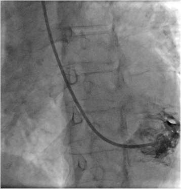
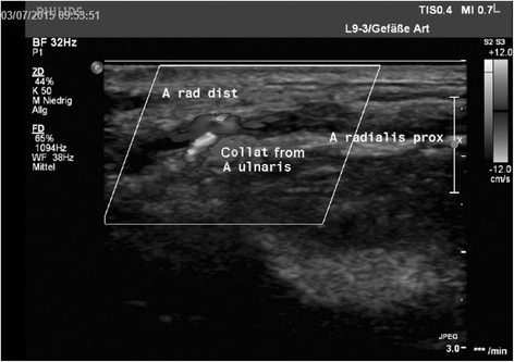
Methods and results: We evaluated the transradial access using a 7.5 F sheathless multipurpose guiding catheter to obtain EMBs from the left ventricle (LV). 18 patients were included. The transradial success rate was 100% (18/18). There were no periprocedural cardiac complications. Immediate post-procedural ambulation could be achieved in all patients. Although radial artery pulse was confirmed by ultrasonic vascular Doppler after removal of the guide in 100% (18/18) of the patients, 50% (9/18) of the patients showed occlusion of the radial artery RAO) by duplex sonography proximal to the access site. 33% (3/9) of the patients in the RAO group and 11,1% (1/9) of the patients in the patent radial artery (RAP) group, respectively, experienced mild pain after the procedure in the right lower arm. Colour Doppler ultrasonography of the right radial artery performed 24 h after the procedure revealed radial occlusion in 50% (9/18) of the patients. The diameter of the radial artery was significantly smaller in the RAO group (p = 0,034), peak systolic velocity (PSV) of the right ulnar artery was significantly higher in the RAO group (p = 0.012). Peak systolic velocity of the opposite radial artery was significantly lower in the RAO group (p = 0,045). Gender, sex, diabetes, radial artery inner diameter ≤2.5 mm and lower peak systolic velocity of < 50 cm/s are predictors of RAO.
Conclusion: The present study demonstrates the safety and efficacy of a transradial access for EMB using a highly hydrophilic sheathless guiding catheter.
Keywords: Endomyocardial biopsy; Sheathless guiding catheter; Transradial approach.
Figures


References
-
- Caforio AL, Pankuweit S, Arbustini E, Basso C, Gimeno-Blanes J, Felix SB, Fu M, Helio T, Heymans S, Jahns R, et al. Current state of knowledge on aetiology, diagnosis, management, and therapy of myocarditis: a position statement of the European Society of Cardiology Working Group on Myocardial and Pericardial Diseases. Eur Heart J. 2013;34(33):2636–48. doi: 10.1093/eurheartj/eht210. - DOI - PubMed
-
- Cooper LT, Baughman KL, Feldman AM, Frustaci A, Jessup M, Kuhl U, Levine GN, Narula J, Starling RC, Towbin J, et al. The role of endomyocardial biopsy in the management of cardiovascular disease: a scientific statement from the American Heart Association, the American College of Cardiology, and the European Society of Cardiology. Endorsed by the Heart Failure Society of America and the Heart Failure Association of the European Society of Cardiology. J Am Coll Cardiol. 2007;50(19):1914–31. doi: 10.1016/j.jacc.2007.09.008. - DOI - PubMed
-
- Escher F, Lassner D, Kuhl U, Gross U, Westermann D, Poller W, Skurk C, Weitmann K, Hoffmann W, Tschope C, et al. Analysis of endomyocardial biopsies in suspected myocarditis--diagnostic value of left versus right ventricular biopsy. Int J Cardiol. 2014;177(1):76–8. doi: 10.1016/j.ijcard.2014.09.071. - DOI - PubMed
-
- Holzmann M, Nicko A, Kuhl U, Noutsias M, Poller W, Hoffmann W, Morguet A, Witzenbichler B, Tschope C, Schultheiss HP, et al. Complication rate of right ventricular endomyocardial biopsy via the femoral approach: a retrospective and prospective study analyzing 3048 diagnostic procedures over an 11-year period. Circulation. 2008;118(17):1722–8. doi: 10.1161/CIRCULATIONAHA.107.743427. - DOI - PubMed
Publication types
MeSH terms
LinkOut - more resources
Full Text Sources
Other Literature Sources
Medical

