. 2016 Dec 9;17(1):250.
doi: 10.1186/s13059-016-1109-7.

Tracing the origin of disseminated tumor cells in breast cancer using single-cell sequencing

Affiliations

Tracing the origin of disseminated tumor cells in breast cancer using single-cell sequencing

Jonas Demeulemeester et al. Genome Biol. .

Abstract

Background: Single-cell micro-metastases of solid tumors often occur in the bone marrow. These disseminated tumor cells (DTCs) may resist therapy and lay dormant or progress to cause overt bone and visceral metastases. The molecular nature of DTCs remains elusive, as well as when and from where in the tumor they originate. Here, we apply single-cell sequencing to identify and trace the origin of DTCs in breast cancer.

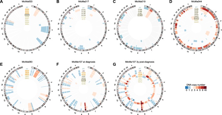
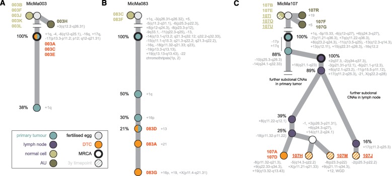
Results: We sequence the genomes of 63 single cells isolated from six non-metastatic breast cancer patients. By comparing the cells' DNA copy number aberration (CNA) landscapes with those of the primary tumors and lymph node metastasis, we establish that 53% of the single cells morphologically classified as tumor cells are DTCs disseminating from the observed tumor. The remaining cells represent either non-aberrant "normal" cells or "aberrant cells of unknown origin" that have CNA landscapes discordant from the tumor. Further analyses suggest that the prevalence of aberrant cells of unknown origin is age-dependent and that at least a subset is hematopoietic in origin. Evolutionary reconstruction analysis of bulk tumor and DTC genomes enables ordering of CNA events in molecular pseudo-time and traced the origin of the DTCs to either the main tumor clone, primary tumor subclones, or subclones in an axillary lymph node metastasis.

Conclusions: Single-cell sequencing of bone marrow epithelial-like cells, in parallel with intra-tumor genetic heterogeneity profiling from bulk DNA, is a powerful approach to identify and study DTCs, yielding insight into metastatic processes. A heterogeneous population of CNA-positive cells is present in the bone marrow of non-metastatic breast cancer patients, only part of which are derived from the observed tumor lineages.

Keywords: Disseminated tumor cells; Intra-tumor genetic heterogeneity; Metastasis; Phylogeny; Single-cell sequencing.


Figures

Fig. 1
Fig. 1
DNA copy number profiles of breast tumors and bone marrow-derived single cells. ag Per patient profiles are shown as concentric circles inside the circular human karyogram. Total (clonal) copy number is represented as a heat map from blue to red as indicated. Tumor samples and single cells are labeled on the corresponding circles and are color-coded: primary tumor bulk (PT) in cyan, lymph node bulk (LN) in purple, DTCs in orange, aberrant cells of unknown origin in dark green, and normal cells in green. Cells isolated after MOPC 21 isotype control staining are boxed. Panels are shown for patient MicMa003 (a), MicMa017 (b), MicMa019 (c), MicMa044 (d), MicMa083 (e), and MicMa107 at the time of diagnosis (f) and 3 years post-diagnosis (g)
Fig. 2
Fig. 2
Genotyping of single nucleotide variants from bulk tumor exome sequences in the single-cell sequences. a, b Heat maps per tumor, where each row represents either a single somatic substitution called on the corresponding bulk exome and the matched blood (a) or a random heterozygous germline SNP (20 total) (b), and columns represent the different single cell (DTC, normal, or AU) or exome datasets obtained for that tumor. Tile colors reflect the detection of the variant allele (orange), of the reference allele only (blue), or whether there was no coverage at that position (white). Only DTCs, but no normal or AU cells, share mutations with the tumor. A single mutation shared between normal cell 107B and the tumor of MicMa107 was later confirmed as a missed heterozygous germline variant (indicated with an asterisk). For clarity, loci with zero coverage in all of the single cells of that patient are omitted. c Modeled probability of observing an at least equally extreme pattern of somatic reference and variant alleles for that cell only through false positives (i.e., the cell derives from another lineage and has none of the tumor’s somatic mutations) or false negatives (the cell derives from the tumor and contains these specific somatic mutations). Model results are encoded as heat maps of − log10(p)
Fig. 3
Fig. 3
Aberrant cells of unknown origin show recurrent aberrations and correlate with age. a Gains (red), losses (blue), and copy neutral loss of heterozygosity (cnLOH, green) events observed in the AUs of the different patients. b Linear regression analysis of the fraction of aberrant non-DTCs versus patient age. Shaded areas represent the regression 95% confidence interval and error bars the standard error of the estimated proportion. All cells (doublet constituents and quality control-failed) were taken into account to estimate the fractions
Fig. 4
Fig. 4
Tracing the origins of DTCs in the breast cancer phylogenetic trees. Copy number-based phylogenetic trees drawn up for patients MicMa003 (a), MicMa083 (b), and MicMa107 (c). Nodes in the trees correspond to (sub)clones and are color-coded based on their type as indicated (primary or lymph node, DTC, AU, or normal cell). Grey nodes are not observed directly but can be inferred from the data. Nodes are annotated with their specific CNA event and, where possible, with their estimated cancer cell fraction (the percentage of tumor cells containing the indicated aberration). Within the tumors, branch lengths reflect differences in cancer cell fraction. The most recent common ancestor (MRCA) in each bulk sample is indicated with a thicker stroke. For MicMa107, single cells isolated 3 years post-diagnosis are represented as striped nodes. DTC 107M has undergone a whole-genome duplication (WGD)

Comment in

  • A case of mistaken identity.
    Shalek AK. Shalek AK. Sci Transl Med. 2017 Jan 4;9(371):eaal4988. doi: 10.1126/scitranslmed.aal4988. Sci Transl Med. 2017. PMID: 28053155 No abstract available.

References

    1. Stratton MR, Campbell PJ, Futreal PA. The cancer genome. Nature. 2009;458:719–24. doi: 10.1038/nature07943. - DOI - PMC - PubMed
    1. Yates LR, Campbell PJ. Evolution of the cancer genome. Nat Rev Genet. 2012;13:795–806. doi: 10.1038/nrg3317. - DOI - PMC - PubMed
    1. Baca SC, Prandi D, Lawrence MS, Mosquera JM, Romanel A, Drier Y, Park K, Kitabayashi N, MacDonald TY, Ghandi M, et al. Punctuated evolution of prostate cancer genomes. Cell. 2013;153:666–77. doi: 10.1016/j.cell.2013.03.021. - DOI - PMC - PubMed
    1. Nik-Zainal S, Van Loo P, Wedge DC, Alexandrov LB, Greenman CD, Lau KW, Raine K, Jones D, Marshall J, Ramakrishna M, et al. The life history of 21 breast cancers. Cell. 2012;149:994–1007. doi: 10.1016/j.cell.2012.04.023. - DOI - PMC - PubMed
    1. Shah SP, Roth A, Goya R, Oloumi A, Ha G, Zhao Y, Turashvili G, Ding J, Tse K, Haffari G, et al. The clonal and mutational evolution spectrum of primary triple-negative breast cancers. Nature. 2012;486:395–9. - PMC - PubMed

Publication types

MeSH terms

Substances