. 2016 Dec 8;14(1):30.
doi: 10.1186/s12964-016-0153-y.

HtrA-mediated E-cadherin cleavage is limited to DegP and DegQ homologs expressed by gram-negative pathogens

Affiliations

HtrA-mediated E-cadherin cleavage is limited to DegP and DegQ homologs expressed by gram-negative pathogens

Carmen M Abfalter et al. Cell Commun Signal. .

Abstract

Background: The serine proteases HtrA/DegP secreted by the human gastrointestinal pathogens Helicobacter pylori (H. pylori) and Campylobacter jejuni (C. jejuni) cleave the mammalian cell adhesion protein E-cadherin to open intercellular adhesions. A wide range of bacteria also expresses the HtrA/DegP homologs DegQ and/or DegS, which significantly differ in structure and function.

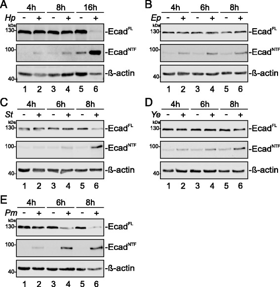
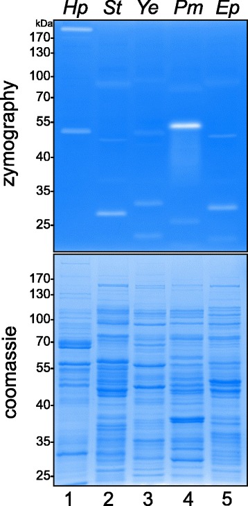
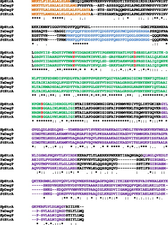
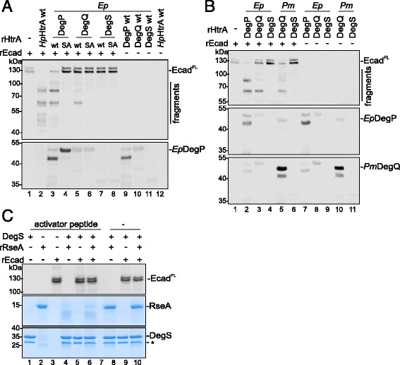
Methods: E-cadherin shedding was investigated in infection experiments with the Gram-negative pathogens H. pylori, enteropathogenic Escherichia coli (EPEC), Salmonella enterica subsp. Enterica (S. Typhimurium), Yersinia enterocolitica (Y. enterocolitica), and Proteus mirabilis (P. mirabilis), which express different combinations of HtrAs. Annotated wild-type htrA/degP, degQ and degS genes were cloned and proteolytically inactive mutants were generated by a serine-to-alanine exchange in the active center. All HtrA variants were overexpressed and purified to compare their proteolytic activities in casein zymography and in vitro E-cadherin cleavage experiments.

Results: Infection of epithelial cells resulted in a strong E-cadherin ectodomain shedding as reflected by the loss of full length E-cadherin in whole cell lysates and formation of the soluble 90 kDa extracellular domain of E-cadherin (NTF) in the supernatants of infected cells. Importantly, comparing the caseinolytic and E-cadherin cleavage activities of HtrA/DegP, DegQ and DegS proteins revealed that DegP and DegQ homologs from H. pylori, S. Typhimurium, Y. enterocolitica, EPEC and P. mirabilis, but not activated DegS, cleaved E-cadherin as a substrate in vitro.

Conclusions: These data indicate that E-cadherin cleavage is confined to HtrA/DegP and DegQ proteins representing an important prevalent step in bacterial pathogenesis.

Keywords: DegP; DegQ; E-cadherin; HtrA.


Figures

Fig. 1
Fig. 1
E-cadherin cleavage during infection with Gram-negative pathogens. Human epithelial cells were infected with (a) H. pylori (Hp) at a MOI 100, (b) EPEC (Ep) at a MOI 5, (c) S. Typhimurium (St) at a MOI 5, (d) Y. enterocolitica (Ye) at a MOI 50 and (e) P. mirabilis (Pm) at a MOI 2. Different MOIs were chosen after careful titration of infection doses to minimize bacterial overgrowth during infection. After indicated time periods, cells were lysed and full length E-cadherin (EcadFL) was detected by Western blot analyses using an antibody against the intracellular domain. Aliquots of supernatants were analyzed for the soluble extracellular E-cadherin fragment (EcadNTF) using an antibody against the extracellular domain. β-actin served as a loading control
Fig. 2
Fig. 2
Active proteases expressed by pathogens. H. pylori (Hp), EPEC (Ep), S. Typhimurium (St), Y. enterocolitica (Ye) and P. mirabilis (Pm) were sonicated and protein lysates were analyzed by casein zymography (upper panel). As a control, proteins were separated by SDS PAGE followed by coomassie staining to show equal protein loading (lower panel)
Fig. 3
Fig. 3
Sequence alignment of the different HtrA/DegP proteins. Signal peptides (orange), proteolytic domains (green) containing the catalytic triad (red) and two PDZ domains (purple) of H. pylori HtrA (HpHtrA), Y. enterocolitica DegP (YeDegP), EPEC DegP (EpDegP) and S. Typhimurium HtrA (StHtrA) are indicated. The LA loop region is highlighted in blue
Fig. 4
Fig. 4
Recombinant HtrA’s/DegP’s are proteolytically active and cleave E-cadherin in vitro. a Domain architecture of HtrA/DegP, DegQ and DegS proteins. SP, signal peptide (orange); protease domain (green); PDZ domains (purple); TMD, transmembrane domain (red). b The proteolytic activity of recombinant HtrA/DegP (rHtrA) wildtype proteins (wt) of H. pylori (Hp), S. Typhimurium (St), Y. enterocolitica (Ye), EPEC (Ep) and DegQ of P. mirabilis (Pm) was analyzed by casein zymography and compared to their corresponding inactive mutants (SA) (upper panel). Coomassie-stained SDS PAGEs demonstrated equal protein loading (lower panel). Self-processed proteins (black asterisks) exhibiting proteolytic activity (white asterisks) are indicated. c Recombinant HtrAs/DegPs (wt) were investigated in in vitro cleavage assays using E-cadherin (E-cadFL) as a substrate and compared with the corresponding inactive variants (SA) as a control. Fragments of E-cadherin were detected using an antibody recognizing the extracellular domain domain. HtrA/DegP proteins were detected using corresponding polyclonal antibodies
Fig. 5
Fig. 5
DegP and DegQ, but not DegS cleave E-cadherin in vitro. a DegP, DegQ and DegS wildtype (wt) of EPEC (Ep) and the corresponding inactive mutants (SA) were tested in in vitro cleavage assays using E-cadherin (rEcad) as a substrate (upper panel). EpDegPwt and EpDegPSA were detected using anti-EpDegP antibody (lower panel). b The E-cadherin-cleavage activity of EPEC (Ep) DegP, DegQ and DegS was compared with the activity of P. mirabilis (Pm) DegQ and DegS. EpDegP and PmDegQ were detected using polyclonal antibodies. c The selective activity of EpDegS was shown in in vitro cleavage experiments using 7 μg EpDegS and 9 μg recombinant RseA (rRseA) as a substrate. To stimulate the activity of EpDegS, 100 μM YFF activator peptide or equal amounts of diluent (−) were added as indicated. 300 ng rEcad was included in the reactions where indicated. Aliquots of samples were analyzed by Western blotting to detect E-cadherin (upper panel) and the remaining sample was separated by SDS PAGE following coomassie staining to detect the degradation of RseA (middle panel) and EpDegS proteins (lower panel). The asterisk (*) indicates GST protein co-purified with the EpDegS protein

References

    1. Clausen T, Kaiser M, Huber R, Ehrmann M. HTRA proteases: regulated proteolysis in protein quality control. Nat Rev Mol Cell Biol. 2011;12(3):152–162. doi: 10.1038/nrm3065. - DOI - PubMed
    1. Skorko-Glonek J, Zurawa-Janicka D, Koper T, Jarzab M, Figaj D, Glaza P, Lipinska B. HtrA protease family as therapeutic targets. Curr Pharm Des. 2013;19(6):977–1009. doi: 10.2174/1381612811319060003. - DOI - PubMed
    1. Hansen G, Hilgenfeld R. Architecture and regulation of HtrA-family proteins involved in protein quality control and stress response. Cell Mol Life Sci. 2013;70(5):761–775. doi: 10.1007/s00018-012-1076-4. - DOI - PMC - PubMed
    1. Sawa J, Heuck A, Ehrmann M, Clausen T. Molecular transformers in the cell: lessons learned from the DegP protease-chaperone. Curr Opin Struct Biol. 2010;20(2):253–258. doi: 10.1016/j.sbi.2010.01.014. - DOI - PubMed
    1. Krojer T, Sawa J, Schafer E, Saibil HR, Ehrmann M, Clausen T. Structural basis for the regulated protease and chaperone function of DegP. Nature. 2008;453(7197):885–890. doi: 10.1038/nature07004. - DOI - PubMed

Publication types

MeSH terms

LinkOut - more resources