

Influence of Rack Design and Disease Prevalence on Detection of Rodent Pathogens in Exhaust Debris Samples from Individually Ventilated Caging Systems
- PMID: 27931317
- PMCID: PMC5113880
Influence of Rack Design and Disease Prevalence on Detection of Rodent Pathogens in Exhaust Debris Samples from Individually Ventilated Caging Systems
Abstract
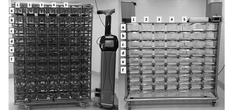
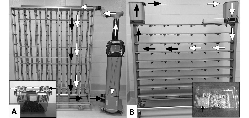
Sampling of bedding debris within the exhaust systems of ventilated racks may be a mechanism for detecting murine pathogens in colony animals. This study examined the effectiveness of detecting pathogens by PCR analysis of exhaust debris samples collected from ventilated racks of 2 different rack designs, one with unfiltered air flow from within the cage to the air-exhaust pathway, and the other had a filter between the cage and the air-exhaust pathway. For 12 wk, racks were populated with either 1 or 5 cages of mice (3 mice per cage) infected with one of the following pathogens: mouse norovirus (MNV), mouse parvovirus (MPV), mouse hepatitis virus (MHV), Helicobacter spp., Pasteurella pneumotropica, pinworms, Entamoeba muris, Tritrichomonas muris, and fur mites. Pathogen shedding by infected mice was monitored throughout the study. In the filter-containing rack, PCR testing of exhaust plenums yielded negative results for all pathogens at all time points of the study. In the rack with open air flow, pathogens detected by PCR analysis of exhaust debris included MHV, Helicobacter spp., P. pneumotropica, pinworms, enteric protozoa, and fur mites; these pathogens were detected in racks housing either 1 or 5 cages of infected mice. Neither MPV nor MNV was detected in exhaust debris, even though prolonged viral shedding was confirmed. These results demonstrate that testing rack exhaust debris from racks with unfiltered air flow detected MHV, enteric bacteria and parasites, and fur mites. However, this method failed to reliably detect MNV or MPV infection of colony animals.
Figures
References
-
- Artwohl JE, Cera LM, Wright MF, Medina LV, Kim LJ. 1994. The efficacy of a dirty-bedding sentinel system for detecting Sendai virus infection in mice: a comparison of clinical signs and seroconversion. Lab Anim Sci 44:73–75. - PubMed
-
- Brielmeier M, Mahabir E, Needham JR, Lengger C, Wilhelm P, Schmidt J. 2006. Microbiologic monitoring of laboratory mice and biocontainment in individually ventilated cages: a field study. Lab Anim 40:247–260. - PubMed
-
- Compton SR, Homberger FR, Paturzo FX, Clark JM. 2004. Efficacy of 3 microbiologic monitoring methods in a ventilated cage rack. Comp Med 54:382–392. - PubMed
-
- Dillehay DL, Lehner ND, Huerkamp MJ. 1990. The effectiveness of a microisolator cage system and sentinel mice for controlling and detecting MHV and Sendai virus infections. Lab Anim Sci 40:367–370. - PubMed
MeSH terms
LinkOut - more resources
Full Text Sources
Miscellaneous