Review
. 2017 Aug;43(5):909-932.
doi: 10.1016/j.burns.2016.11.014. Epub 2016 Dec 5.

Thermal injury of skin and subcutaneous tissues: A review of experimental approaches and numerical models

Affiliations
Review

Thermal injury of skin and subcutaneous tissues: A review of experimental approaches and numerical models

Hanglin Ye et al. Burns. 2017 Aug.

Abstract

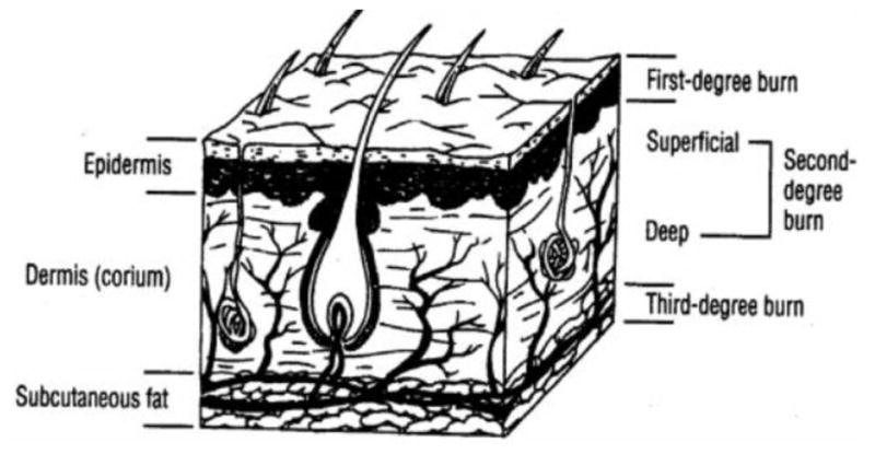
Thermal injury to skin and subcutaneous tissue is common in both civilian and combat scenarios. Understanding the change in tissue morphologies and properties and the underlying mechanisms of thermal injury are of vital importance to clinical determination of the degree of burn and treatment approach. This review aims at summarizing the research involving experimental and numerical studies of skin and subcutaneous tissue subjected to thermal injury. The review consists of two parts. The first part deals with experimental studies including burn protocols and prevailing imaging approaches. The second part deals with existing numerical models for burns of tissue and related computational simulations. Based on this review, we conclude that though there is literature contributing to the knowledge of the pathology and pathogenesis of tissue burn, there is scant quantitative information regarding changes in tissue properties including mechanical, thermal, electrical and optical properties as a result of burns that are linked to altered tissue morphology.

Keywords: Burn imaging; Burning experiment protocols; Review; Soft tissue burns; Thermal injury mechanisms; Thermal injury simulations.


Conflict of interest statement

Conflicts of interest: none

Figures

Fig. 1
Fig. 1
Diagram for skin anatomy showing different degrees of burns, adopted with permission from [12].
Fig. 2
Fig. 2
(×400) Histologic images of porcine (left) and human (right) skin of severe first degree burns, adopted with permission from [46].
Fig. 3
Fig. 3
(×400) Histologic images of porcine (left) and human (right) skin of early second degree burns, adopted with permission from [46].
Fig. 4
Fig. 4
(×400) Histologic images of third degree burnt porcine tissue 24 hours post-burn, with (left) and without (right) coagulated dermis, adopted with permission from [46].
Fig. 5
Fig. 5
(×400) Histologic images of third degree burnt porcine tissue, 72 hours post-burn, adopted with permission from [46].
Fig. 6
Fig. 6
(×85) Histologic images of extremely burnt porcine tissue, adopted with permission from [46]
Fig. 7
Fig. 7
(×1050) Histologic image of blocked vessel 48 hours post-burn, adopted with permission from [3].
Fig. 8
Fig. 8
(×1050) Histologic image of patent vessel, adopted with permission from [3]
Fig. 9
Fig. 9
Laser Doppler imaging at day 0, 1, 3, 5 and 8 post-burn, showing wound healing and increasing perfusion. The healed wound at day 21 post burn is also shown, adopted with permission from [5].
Fig. 10
Fig. 10
Videomicroscopy images of different grades of burn depth, adopted from [67].
Fig. 11
Fig. 11
Videomicroscopy findings of capillary patterns corresponding to different burn depth. SDB refers to ‘superficial dermal burns’, while DDB refers to ‘deep dermal burns’, adopted with permission from [68].
Fig. 12
Fig. 12
Orthogonal polarization spectral images of A) normal skin, with visible dermal capillaries and individual red blood cells; (B) superficial thermal burn, a resemblance to the normal skin; (C) deep thermal burn, with visible larger coagulated thrombosis, adopted with permission from [69].
Fig. 13
Fig. 13
RCM images of stratum granulosum layer of skin. (a) healthy tissue with cells appearing as dark nuclei (b) superficial burnt tissue with larger cells (c) superficial partial thickness burnt tissue with destroyed cell structure (d) deep partial thickness burnt tissue with cells appearing in a dark shadow Figure is adopted with permission from [72].
Fig. 14
Fig. 14
True-color image (left) and false-color image (right) from MSI, adopted with permission from [4]. Blue color indicates deep partial thickness; green indicates deep-dermal thickness and red indicates full-thickness burns.
Fig. 15
Fig. 15
Comparison of images from LSI and LDI. (a) photograph of a hypertrophic from LSI CCD camera; (b) LSI image showing tissue perfusion on the same site; (c) Moor LDI total reflected light intensity map of the same site; (d) Moor LDI image showing perfusion of the same site. Figure is adopted with permission from [96].
Fig. 16
Fig. 16
Time-temperature threshold for thermal injury and second degree burn, replotted with permission from [110].

References

    1. American Burn Associations. Burn Incidence Fact Sheet. 2016.
    1. Deitch EA, Wheelahan TM, Rose MP, Clothier J, Cotter J. Hypertrophic burn scars: analysis of variables. J Trauma. 1983;23(10):895–898. - PubMed
    1. Watts AMI, Tyler MPH, Perry ME, Roberts AHN, McGrouther DA. Burn depth and its histological measurement. Burns. 2001;27(2):154–160. - PubMed
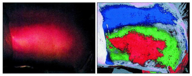
    1. Eisenbeiß W, Marotz J, Schrade JP. Reflection-optical multispectral imaging method for objective determination of burn depth. Burns. 1999;25(8):697–704. - PubMed
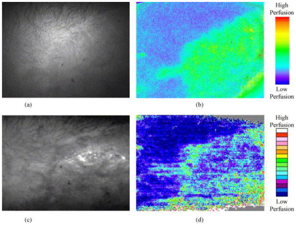
    1. Hoeksema H, Van de Sijpe K, Tondu T, Hamdi M, Van Landuyt K, Blondeel P, Monstrey S. Accuracy of early burn depth assessment by laser Doppler imaging on different days post burn. Burns. 2009;35(1):36–45. - PubMed