Review
. 2017 Apr;27(4):266-275.
doi: 10.1016/j.tcb.2016.11.004. Epub 2016 Dec 5.

Bax and Bak Pores: Are We Closing the Circle?

Affiliations
Review

Bax and Bak Pores: Are We Closing the Circle?

Katia Cosentino et al. Trends Cell Biol. 2017 Apr.

Abstract

Bax and its homolog Bak are key regulators of the mitochondrial pathway of apoptosis. On cell stress Bax and Bak accumulate at distinct foci on the mitochondrial surface where they undergo a conformational change, oligomerize, and mediate cytochrome c release, leading to cell death. The molecular mechanisms of Bax and Bak assembly and mitochondrial permeabilization have remained a longstanding question in the field. Recent structural and biophysical studies at several length scales have shed light on key aspects of Bax and Bak function that have shifted how we think this process occurs. These discoveries reveal an unexpected molecular mechanism in which Bax (and likely Bak) dimers assemble into oligomers with an even number of molecules that fully or partially delineate pores of different sizes to permeabilize the mitochondrial outer membrane (MOM) during apoptosis.

Keywords: Bax; Bcl-2 proteins; apoptosis; cell death; mitochondrial outer membrane permeabilization; pore-forming proteins.


Figures

Figure 1
Figure 1. Model for Bax Activation and Oligomerization at the Membrane.
Proteins are illustrated in their surface representations. In its soluble form, Bax (cyan) retains a globular conformation where its transmembrane domain (∝9 helix, blue) is kept in the Bax hydrophobic groove (∝2–∝5). On activation by Bcl-2 homology (BH)3-only proteins (yellow), Bax undergoes a series of conformational changes displacing ∝9 from the groove, which inserts in the membrane (gray), and dissociating in a core (∝2–∝5) and a latch (∝6–∝9) domain by unfolding of helices ∝5 and ∝6. Following activation, Bax engages in BH3-in-groove dimerization with another activated Bax molecule (light green) while displacing the BH3-only activator from the hydrophobic groove. This leads to the formation of a symmetric dimer. Further oligomerization proceeds by the addition of other dimer units via a second, as-yet unclear, interaction site.
Figure 2
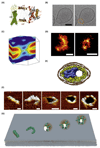
Figure 2. Evidence of Bax Pore Formation.
(A) Clamp model of the Bax dimer at the membrane. The dimerization domain is at the edge of a membrane pore induced by Bax while ∝6 helices are located on opposite sides of the membrane. The orientation of the C-terminal ∝9 helices is unclear. Reproduced, with permission, from [18]. (B) Cryoelectron microscopy (cryo-EM) image of gold-labeled Bax delineating a pore in the membrane of a vesicle. In the corresponding image, the pore is outlined in yellow and Bax in orange for visual purposes. Bar, 40 nm. Reproduced, with permission, from [51]. (C) X-ray diffraction structure of a pore formed by the ∝5 peptide of Bax. Yellow-to-red regions represent high electron density of Br atoms localized at the lipid chains. Electron density is low (blue regions) at the pore rim and outside the membrane. Reproduced, with permission, from [45]. (D) Arc and ring structures of GFP-Bax wild type reconstructed by super-resolution single-molecule localization microscopy. Bars, 100 nm. Reproduced, with permission, from [48]. (E) Model of nanodisk structure (yellow) containing a single unit of BAX/BID peptide (blue surface) based on cryo-EM data [50]. Reproduced, with permission, from [82]. (F) Atomic force microscopy (AFM) images of structures made by Bax (yellow protrusions) on supported lipid bilayers (brown areas) showing (left to right) a line, arcs of different lengths, and a ring. The black area in the last three images is representative of a hole in the membrane. Bax molecules partially or completely line the pore rim. Bars, 50 nm. (G) Models for Bax (cyan) structures, including (left to right) lines, arcs, and rings, leading to pore formation in the membrane (gray).
Figure 3
Figure 3. Regulation of Bax Activity by Other Bcl-2 Proteins.
(A) Under healthy conditions Bax (cyan) associates with the mitochondrial outer membrane (MOM) (gray) but is continuously retrotranslocated to the cytosol by prosurvival Bcl-2 proteins. (B) During apoptosis Bcl-2 homology (BH)3-only proteins break this steady state by inhibiting the action of prosurvival Bcl-2 proteins and activating Bax. Insertion of ∝9 (blue) in the membrane and further conformational changes allow Bax oligomerization and induce MOM permeabilization (MOMP).

References

    1. Strasser A, et al. Deciphering the rules of programmed cell death to improve therapy of cancer and other diseases. EMBO J. 2011;30:3667–3683. - PMC - PubMed
    1. Czabotar PE, et al. Control of apoptosis by the BCL-2 protein family: implications for physiology and therapy. Nat Rev Mol Cell Biol. 2014;15:49–63. - PubMed
    1. Delbridge ARD, et al. Thirty years of BCL-2: translating cell death discoveries into novel cancer therapies. Nat Rev Cancer. 2016;16:99–109. - PubMed
    1. García-Sáez AJ. The secrets of the Bcl-2 family. Cell Death Differ. 2012;19:1733–1740. - PMC - PubMed
    1. Li P, et al. Cytochrome c and dATP-dependent formation of Apaf-1/caspase-9 complex initiates an apoptotic protease cascade. Cell. 1997;91:479–489. - PubMed

Publication types

Substances