Review
. 2017 Mar 1:617:68-83.
doi: 10.1016/j.abb.2016.12.003. Epub 2016 Dec 6.

Peroxiredoxin 6 in the repair of peroxidized cell membranes and cell signaling

Affiliations
Review

Peroxiredoxin 6 in the repair of peroxidized cell membranes and cell signaling

Aron B Fisher. Arch Biochem Biophys. .

Abstract

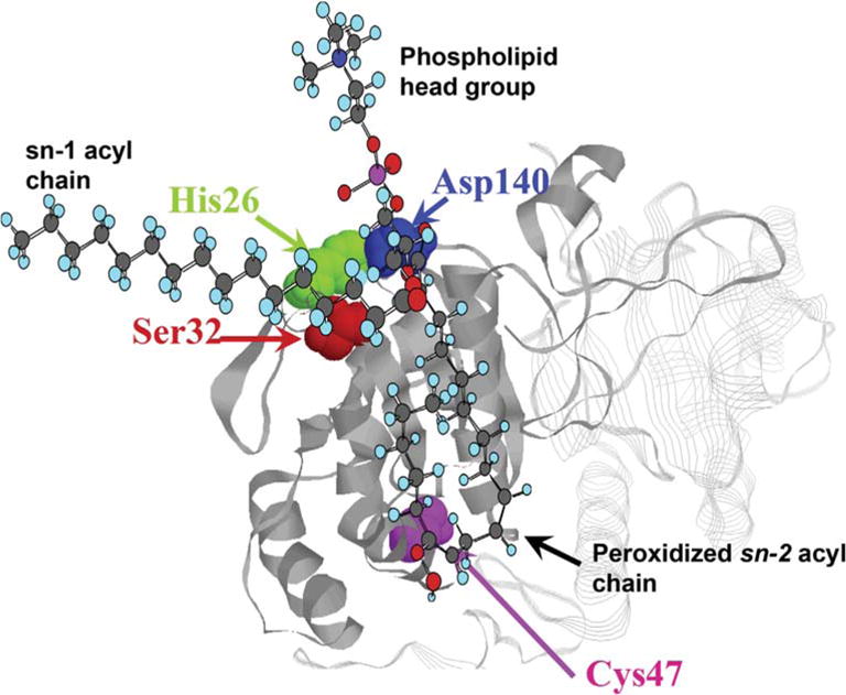
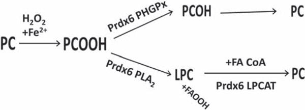
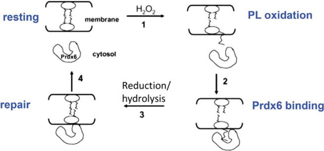
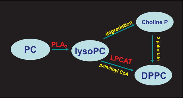
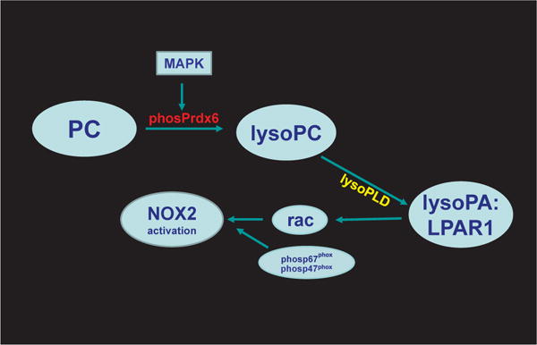
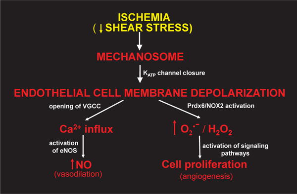
Peroxiredoxin 6 represents a widely distributed group of peroxiredoxins that contain a single conserved cysteine in the protein monomer (1-cys Prdx). The cys when oxidized to the sulfenic form is reduced with glutathione (GSH) catalyzed by the π isoform of GSH-S-transferase. Three enzymatic activities of the protein have been described:1) peroxidase with H2O2, short chain hydroperoxides, and phospholipid hydroperoxides as substrates; 2) phospholipase A2 (PLA2); and 3) lysophosphatidylcholine acyl transferase (LPCAT). These activities have important physiological roles in antioxidant defense, turnover of cellular phospholipids, and the generation of superoxide anion via initiation of the signaling cascade for activation of NADPH oxidase (type 2). The ability of Prdx6 to reduce peroxidized cell membrane phospholipids (peroxidase activity) and also to replace the oxidized sn-2 fatty acyl group through hydrolysis/reacylation (PLA2 and LPCAT activities) provides a complete system for the repair of peroxidized cell membranes.

Keywords: Anti-oxidant defense; Lysophospholipid acyl transferase; NADPH oxidase; Phospholipase A(2); Phospholipid hydroperoxide glutathione peroxidase; Phospholipid remodeling.


Figures

Fig. 1
Fig. 1. Amino acid sequence for mouse Prdx6
The full length sequence is shown. The consensus sequences are indicated for peroxidase and LPCAT activities, the PLA2 and peroxidase catalytic triads, the lipase motif, the motif (LBTM) for targeting of Prdx6 to the lamellar bodies, the hydrophobic surface (HS) for dimerization, the SP-A binding sequence, and the Prdx6 phosphorylation site.
Fig. 2
Fig. 2. Mechanism for Prdx6 peroxidase activity and its resolution
Clockwise from upper left. (A) In solution, Prdx6 exists as a homodimer through hydrophobic interactions surrounding the L145-L148 residues; the reduced peroxidatic sulfhydryl (SH) is at the bottom of a pocket. (B) Reduction of H2O2 (peroxidase activity) results in oxidation of the Prdx6 sulfhydryl to the sulfenic form (SOH) leading to dissociation of the homodimer. Interaction of the oxidized monomer of Prdx6 with GSH-loaded πGST results in: (C) heterodimerization via the L145-L148 hydrophobic interface of Prdx6 and glutathionylation of Prdx6 (SSG), followed by: (D) dissociation of the glutathionylated Prdx6 and πGST heterodimer. Interaction of the glutathionylated monomer with free GSH restores its normally reduced state allowing reformation of the homodimer.
Fig. 3
Fig. 3. Reaction scheme for Prdx6 peroxidase activity with proposed formation of a sulfenylamide intermediate
Interaction of Prdx6 with H2O2 generates the sulfenic state (Prdx6-SOH) of the reactive cysteine. The sulfenic cys is susceptible to further (irreversible) oxidation to the sulfinic form (Prdx6-SOOH). The formation of a reversible sulfenylamide (Prdx6-SN) could stabilize the sulfenic state and protect the protein from irreversible oxidation (see text). Interaction of the Prdx6 sulfenylamide with πGST:GSH results in Prdx6 glutathionylation (Prdx6-SSG) and subsequent resolution by interaction with GSH as described in Fig. 2. GSSG formed during the cycle is reduced by NADPH mediated by GSH reductase to regenerate 2 GSH (not shown).
Fig. 4
Fig. 4. Effect of Prdx6 “knock-out” on hydroperoxide reduction by mouse lung homogenate
Knock-out of Prdx6 (Prdx6−/−) has only a minor effect on the GSH-mediated reduction of H2O2 (GPx activity, left y-axis) by the lung homogenate but results in a marked decrease in the GSH-mediated reduction of phosphatidylcholine hydroperoxide (PHGPx activity, right y-axis). Knock-out of GPx1 (GPx1−/−) resulted in the inverse with a large decrease in GPx activity but relatively little effect on PHGPx activity. PCOOH, phosphatidylcholine hydroperoxide. Reprinted from Ref. [64] with permission.
Fig. 5
Fig. 5. Model for phospholipid-associated activities of Prdx6
The model is based on the crystal structure of oxidized human Prdx6 [43]. Prdx6 binds to oxidized phosphatidylcholine at the lipid binding region associated with the lipase motif in the protein. We propose that the peroxidized sn-2 acyl chain inserts into the pocket containing the peroxidatic cys and is positioned at the surface-expressed PLA2 catalytic triad (32S-26H-140D). The sn-1 acyl chain and the phospholipid head group remain outside of the pocket. Thus, Prdx6 can reduce the oxidized acyl moiety (PHGPx activity) or hydrolyze the acyl bond (PLA2 activity) of the peroxidized phospholipid. This complex could be at the cytoplasmic face of a cell membrane at the left (not shown). The hydrophobic sn-1 fatty acyl chain and the hydrophilic head group would be within the membrane while the peroxidized sn-2 fatty acyl chain would be at the membrane surface facilitating interaction with Prdx6. Modified and reprinted with permission from Ref. [78].
Fig. 6
Fig. 6. Role of Prdx6 in the repair of phospholipid hydroperoxides
Phosphatidylcholine (PC) containing an sn-2 unsaturated fatty acid can be oxidized by H2O2 (+Fe2+) to PC hydroperoxide (PCOOH). To restore the normal state, the hydroperoxide can be reduced by the phospholipid hydroperoxide GSH peroxidase (PHGPx) activity of Prdx6 to the PC alcohol that is then reduced to the sulfhydryl. An alternative pathway for removal of PCOOH is hydrolysis at the sn-2 position mediated by Prdx6 PLA2 activity to generate lysoPC plus fatty acyl hydroperoxide; lysoPC can be reacylated with an acyl CoA substrate by the LPCAT activity of Prdx6 to regenerate the reduced phospholipid. Enzymes responsible for reduction of the PC alcohol (PCOH) and fatty acid hydroperoxide (FAOOH) that result from these reactions are not shown. Modified from Ref. [132] and reprinted with permission.
Fig. 7
Fig. 7. Recovery of isolated perfused mouse lungs from lipid peroxidation induced by exposure to t-BOOH
Lungs were exposed to 25 mM (wild type) or 15 mM (mutants) t-BOOH for 1 h followed by 2 h of perfusion with oxidant –free medium. Different concentrations of t-BOOH were used for wild type and mutant lungs to produce equal degrees of oxidant stress during the exposure period. Lung homogenate was analyzed for fluorescence of diphenyl-1-pyrenylphosphine (DPPP), an indicator for phospholipid hydroperoxides [122,163]. The increase of DPPP fluorescence indicates lipid peroxidation. Lungs were from wild type (red), Prdx6 null (blue), C47S Prdx6 knock-in (black), and D140A Prdx6 knock-in (green) mice. The C47S mice do not express the peroxidase activity of Prdx6; the D140A mice do not express the PLA2 activity of Prdx6. Each point is mean for n = 3, the small SE is not shown. Modified and reprinted with permission from Ref. [132].
Fig. 8
Fig. 8. Mechanism for repair of oxidized cell membranes
Prdx6 is cytosolic under normal resting conditions. Oxidation of a membrane phospholipid fatty acyl residue results in its increased hydrophilicity and its ‘flotation’ toward the membrane surface; interaction of Prdx6 with the oxidized lipid results in cell membrane association of the protein. Reduction and/or hydrolysis (plus acyl transferase) of the oxidized fatty acyl moiety ‘repairs’ the cell membrane phospholipid. Prdx6 then dissociates from the membrane to return to the resting state. Prdx6 exists as a dimer in the cytoplasm but only the peroxidatic monomer is shown. Modified from Ref. [33] and reprinted with permission.
Fig. 9
Fig. 9. Pathways for synthesis of dipalmitoyl phosphatidylcholine
These pathways are important for the generation of the major surface active component of the lung surfactant. Lysophosphatidylcholine (lysoPC) is produced by hydrolysis of the sn-2 acyl bond of phosphatidylcholine (PC) through the PLA2 activity of Prdx6. LysoPC can be further degraded by various enzymes; the choline phosphate moiety is conserved and used along with two palmitates for DPPC synthesis by the de novo pathway. sn-1Palmitoyl lysoPC also can be re-acylated with a palmitoyl CoA by the LPCAT activity of Prdx6 (remodeling pathway) to generate DPPC.
Fig. 10
Fig. 10. Pathway for generation of lysophosphatidic acid and the activation of NADPH oxidase type 2 (NOX2)
With an appropriate signal, Prdx6 is phosphorylated via mitogen activated protein kinase (MAPK) activity and binds to phospholipid in the cell membrane. LysoPC that is generated from PC by the PLA2 activity of Prdx6 can be metabolized via lysophospholipase D (lysoPLD) to lysophosphatidic acid (lysoPA). Interaction of lysoPA with its receptor (LPAR type 1) leads to release of rac (rac1 or 2 depending on cell type), one of the cytosolic protein co-factors required for NOX2 activation [146]. Other important co-factors for activation include phosphorylated p47phox and phosphorylated p67phox.
Fig. 11
Fig. 11. Scheme for endothelial mechanotransduction
Decreased shear stress associated with stop of blood flow (ischemia) is sensed by the mechanosome leading to closure of inwardly rectifying KATP channels with resultant cell membrane depolarization, phosphorylation of Prdx6, and activation of NOX2 as shown in Fig. 10. The subsequent generation of O2·/H2O2 results in release of angiogenic factors resulting in cellular proliferation and neovascularization. Cell membrane depolarization also leads to opening of voltage-gated Ca channels (VGCC) followed by Ca2+ influx into the cell and generation of the vasodilating agent NO via endothelial NO synthase (eNOS).

Similar articles

Cited by

References

    1. Joly JS, Bourrat F, Nguyen V, Chourrout D. Ol-Prx 3, a member of an additional class of homeobox genes, is unimodally expressed in several domains of the developing and adult central nervous system of the medaka (Oryzias latipes) Proc Natl Acad Sci U S A. 1997;94:12987–12992. - PMC - PubMed
    1. Shichi H, Demar JC. Non-selenium glutathione peroxidase without gluta-thione S-transferase activity from bovine ciliary body. Experim Eye Res. 1990;50:513–520. - PubMed
    1. Peshenko IV, Novoselov VI, Evdokimov VA, Yu Nikolaev V, Shuvaeva TM, Lipkin VM, Fesenko EE. Novel 28-kDa secretory protein from rat olfactory epithelium. FEBS Lett. 1996;381:12–14. - PubMed
    1. Akiba S, Dodia C, Chen X, Fisher AB. Characterization of acidic Ca(2+)- independent phospholipase A2 of bovine lung. Comp Biochem Physiol B Biochem Mol Biol. 1998;120:393–404. - PubMed
    1. Peshenko IV, Novoselov VI, Evdokimov VA, Nikolaev YV, Kamzalov SS, Shuvaeva TM, Lipkin VM, Fesenko EE. Identification of a 28 kDa secretory protein from rat olfactory epithelium as a thiol-specific antioxidant. Free Radic Biol Med. 1998;25:654–659. - PubMed

MeSH terms