Loss of dopamine D1 receptors and diminished D1/5 receptor-mediated ERK phosphorylation in the periaqueductal gray after spinal cord lesion
- PMID: 27932310
- PMCID: PMC5262528
- DOI: 10.1016/j.neuroscience.2016.11.040
Loss of dopamine D1 receptors and diminished D1/5 receptor-mediated ERK phosphorylation in the periaqueductal gray after spinal cord lesion
Abstract
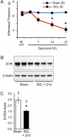
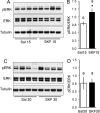
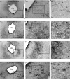
Neuropathic pain resulting from spinal cord injury is often accompanied by maladaptive plasticity of the central nervous system, including the opioid receptor-rich periaqueductal gray (PAG). Evidence suggests that sensory signaling via the PAG is robustly modulated by dopamine D1- and D2-like receptors, but the effect of damage to the spinal cord on D1 and D2 receptor protein expression and function in the PAG has not been examined. Here we show that 21days after a T10 or C6 spinothalamic tract lesion, both mice and rats display a remarkable decline in the expression of D1 receptors in the PAG, revealed by western blot analysis. These changes were associated with a significant reduction in hindpaw withdrawal thresholds in lesioned animals compared to sham-operated controls. We investigated the consequences of diminished D1 receptor levels by quantifying D1-like receptor-mediated phosphorylation of ERK1,2 and CREB, events that have been observed in numerous brain structures. In naïve animals, western blot analysis revealed that ERK1,2, but not CREB phosphorylation was significantly increased in the PAG by the D1-like agonist SKF 81297. Using immunohistochemistry, we found that SKF 81297 increased ERK1,2 phosphorylation in the PAG of sham animals. However, in lesioned animals, basal pERK1,2 levels were elevated and did not significantly increase after exposure to SKF 81297. Our findings provide support for the hypothesis that molecular adaptations resulting in a decrease in D1 receptor expression and signaling in the PAG are a consequence of SCL.
Keywords: SKF 81297; behavior; extracellular signal regulated kinase; hyperalgesia; neuropathic pain; spinal cord injury.
Copyright © 2016 IBRO. Published by Elsevier Ltd. All rights reserved.
Figures


Similar articles
-
Dopamine D1 receptor agonist treatment alleviates morphine-exposure-induced learning and memory impairments.Brain Res. 2019 May 15;1711:120-129. doi: 10.1016/j.brainres.2019.01.020. Epub 2019 Jan 17. Brain Res. 2019. PMID: 30660614
-
Repeated treatments with the D1 dopamine receptor agonist SKF-38393 modulate cell viability via sustained ERK-Bad-Bax activation in dopaminergic neuronal cells.Behav Brain Res. 2019 Jul 23;367:166-175. doi: 10.1016/j.bbr.2019.03.035. Epub 2019 Mar 28. Behav Brain Res. 2019. PMID: 30930179
-
Region-specific deletions of the glutamate transporter GLT1 differentially affect nerve injury-induced neuropathic pain in mice.Glia. 2018 Sep;66(9):1988-1998. doi: 10.1002/glia.23452. Epub 2018 May 3. Glia. 2018. PMID: 29722912
-
The Distinct Functions of Dopaminergic Receptors on Pain Modulation: A Narrative Review.Neural Plast. 2021 Feb 22;2021:6682275. doi: 10.1155/2021/6682275. eCollection 2021. Neural Plast. 2021. PMID: 33688340 Free PMC article. Review.
-
Periaqueductal Gray Sheds Light on Dark Areas of Psychopathology.Trends Neurosci. 2019 May;42(5):349-360. doi: 10.1016/j.tins.2019.03.004. Epub 2019 Apr 4. Trends Neurosci. 2019. PMID: 30955857 Review.
Cited by
-
Neuropathic Pain and Spinal Cord Injury: Management, Phenotypes, and Biomarkers.Drugs. 2023 Jul;83(11):1001-1025. doi: 10.1007/s40265-023-01903-7. Epub 2023 Jun 16. Drugs. 2023. PMID: 37326804 Review.
-
Neuropathic Pain Induced by Spinal Cord Injury from the Glia Perspective and Its Treatment.Cell Mol Neurobiol. 2024 Nov 28;44(1):81. doi: 10.1007/s10571-024-01517-x. Cell Mol Neurobiol. 2024. PMID: 39607514 Free PMC article. Review.
-
Painful stimulation increases spontaneous blink rate in healthy subjects.Sci Rep. 2020 Nov 17;10(1):20014. doi: 10.1038/s41598-020-76804-w. Sci Rep. 2020. PMID: 33203984 Free PMC article.
-
Partially brain effects of injection of human umbilical cord mesenchymal stem cells at injury sites in a mouse model of thoracic spinal cord contusion.Front Mol Neurosci. 2023 Jun 5;16:1179175. doi: 10.3389/fnmol.2023.1179175. eCollection 2023. Front Mol Neurosci. 2023. PMID: 37342099 Free PMC article.
-
Dopamine D1 and D2 receptors mediate analgesic and hypnotic effects of l-tetrahydropalmatine in a mouse neuropathic pain model.Psychopharmacology (Berl). 2019 Nov;236(11):3169-3182. doi: 10.1007/s00213-019-05275-3. Epub 2019 Jun 6. Psychopharmacology (Berl). 2019. PMID: 31172225
References
-
- Acquas E, Pisanu A, Spiga S, Plumitallo A, Zernig G, Di Chiara G. Differential effects of intravenous R,S-(+/−)-3,4-methylenedioxymethamphetamine (MDMA, Ecstasy) and its S(+)- and R(−)-enantiomers on dopamine transmission and extracellular signal regulated kinase phosphorylation (pERK) in the rat nucleus accumbens shell and core. J Neurochem. 2007;102:121–132. - PubMed
-
- Attal N. Neuropathic pain: mechanisms, therapeutic approach, and interpretation of clinical trials. Continuum (Minneap Minn) 2012;18:161–175. - PubMed
-
- Bandler R, Shipley MT. Columnar organization in the midbrain periaqueductal gray: modules for emotional expression? Trends Neurosci. 1994;17:379–389. - PubMed
-
- Baptista-de-Souza D, Di Cesare Mannelli L, Zanardelli M, Micheli L, Nunes-de-Souza RL, Canto-de-Souza A, Ghelardini C. Serotonergic modulation in neuropathy induced by oxaliplatin: effect on the 5HT2C receptor. Eur J Pharmacol. 2014;735:141–9. - PubMed
-
- Beaulieu J-M, Gainetdinov RR. The physiology, signaling, and pharmacology of dopamine receptors. Pharmacol Rev. 2011;63:182–217. - PubMed
Publication types
MeSH terms
Substances
Grants and funding
LinkOut - more resources
Full Text Sources
Other Literature Sources
Medical
Molecular Biology Databases
Research Materials
Miscellaneous

