

Expression of Cav1.3 calcium channel in the human and mouse colon: posttranscriptional inhibition by IFNγ
- PMID: 27932504
- PMCID: PMC5283901
- DOI: 10.1152/ajpgi.00394.2016
Expression of Cav1.3 calcium channel in the human and mouse colon: posttranscriptional inhibition by IFNγ
Abstract
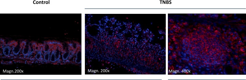
It has been hypothesized that apically expressed L-type Ca2+ channel Cav1.3 (encoded by CACNA1D gene) contributes toward an alternative TRPV6-independent route of intestinal epithelial Ca2+ absorption, especially during digestion when high luminal concentration of Ca2+ and other nutrients limit TRPV6 contribution. We and others have implicated altered expression and activity of key mediators of intestinal and renal Ca2+ (re)absorption as contributors to negative systemic Ca2+ balance and bone loss in intestinal inflammation. Here, we investigated the effects of experimental colitis and related inflammatory mediators on colonic Cav1.3 expression. We confirmed Cav1.3 expression within the segments of the mouse and human gastrointestinal tract. Consistent with available microarray data (GEO database) from inflammatory bowel disease (IBD) patients, mouse colonic expression of Cav1.3 was significantly reduced in trinitrobenzene sulfonic acid (TNBS) colitis. In vitro, IFNγ most potently reduced Cav1.3 expression. We reproduced these findings in vivo with wild-type and Stat1-/- mice injected with IFNγ. The observed effect in Stat1-/- suggested a noncanonical transcriptional repression or a posttranscriptional mechanism. In support of the latter, we observed no effect on the cloned Cav1.3 gene promoter activity and accelerated Cav1.3 mRNA decay rate in IFNγ-treated HCT116 cells. While the relative contribution of Cav1.3 to intestinal Ca2+ absorption and its value as a therapeutic target remain to be established, we postulate that Cav1.3 downregulation in IBD may contribute to the negative systemic Ca2+ balance, to increased bone resorption, and to reduced bone mineral density in IBD patients.
Keywords: calcium absorption; calcium channel; inflammation; interferon; intestine.
Copyright © 2017 the American Physiological Society.
Figures


Similar articles
-
Experimental colitis is associated with transcriptional inhibition of Na+/Ca2+ exchanger isoform 1 (NCX1) expression by interferon γ in the renal distal convoluted tubules.J Biol Chem. 2015 Apr 3;290(14):8964-74. doi: 10.1074/jbc.M114.616516. Epub 2015 Feb 2. J Biol Chem. 2015. PMID: 25648899 Free PMC article.
-
TRPV6 and Cav1.3 Mediate Distal Small Intestine Calcium Absorption Before Weaning.Cell Mol Gastroenterol Hepatol. 2019;8(4):625-642. doi: 10.1016/j.jcmgh.2019.07.005. Epub 2019 Aug 6. Cell Mol Gastroenterol Hepatol. 2019. PMID: 31398491 Free PMC article.
-
Chronic fluoxetine administration increases expression of the L-channel gene Cav1.2 in astrocytes from the brain of treated mice and in culture and augments K(+)-induced increase in [Ca(2+)]i.Cell Calcium. 2014 Mar;55(3):166-74. doi: 10.1016/j.ceca.2014.01.002. Epub 2014 Jan 22. Cell Calcium. 2014. PMID: 24513410
-
L-type CaV1.2 calcium channels: from in vitro findings to in vivo function.Physiol Rev. 2014 Jan;94(1):303-26. doi: 10.1152/physrev.00016.2013. Physiol Rev. 2014. PMID: 24382889 Review.
-
From Gene to Behavior: L-Type Calcium Channel Mechanisms Underlying Neuropsychiatric Symptoms.Neurotherapeutics. 2017 Jul;14(3):588-613. doi: 10.1007/s13311-017-0532-0. Neurotherapeutics. 2017. PMID: 28497380 Free PMC article. Review.
Cited by
-
Intestinal Inflammation Promotes MDL-1+ Osteoclast Precursor Expansion to Trigger Osteoclastogenesis and Bone Loss.Cell Mol Gastroenterol Hepatol. 2022;14(4):731-750. doi: 10.1016/j.jcmgh.2022.07.002. Epub 2022 Jul 12. Cell Mol Gastroenterol Hepatol. 2022. PMID: 35835390 Free PMC article.
-
Calcium physiology, metabolism and supplementation: a glance at patients with ankylosing spondylitis.Reumatologia. 2020;58(5):297-311. doi: 10.5114/reum.2020.100112. Epub 2020 Oct 20. Reumatologia. 2020. PMID: 33227082 Free PMC article. Review.
References
-
- Azizan EA, Poulsen H, Tuluc P, Zhou J, Clausen MV, Lieb A, Maniero C, Garg S, Bochukova EG, Zhao W, Shaikh LH, Brighton CA, Teo AE, Davenport AP, Dekkers T, Tops B, Küsters B, Ceral J, Yeo GS, Neogi SG, McFarlane I, Rosenfeld N, Marass F, Hadfield J, Margas W, Chaggar K, Solar M, Deinum J, Dolphin AC, Farooqi IS, Striessnig J, Nissen P, Brown MJ. Somatic mutations in ATP1A1 and CACNA1D underlie a common subtype of adrenal hypertension. Nat Genet 45: 1055–1060, 2013. doi:10.1038/ng.2716. - DOI - PubMed
-
- Benchimol EI, Ward LM, Gallagher JC, Rauch F, Barrowman N, Warren J, Beedle S, Mack DR. Effect of calcium and vitamin D supplementation on bone mineral density in children with inflammatory bowel disease. J Pediatr Gastroenterol Nutr 45: 538–545, 2007. doi:10.1097/MPG.0b013e3180dca0cc. - DOI - PubMed
-
- Bernstein CN, Seeger LL, Anton PA, Artinian L, Geffrey S, Goodman W, Belin TR, Shanahan F. A randomized, placebo-controlled trial of calcium supplementation for decreased bone density in corticosteroid-using patients with inflammatory bowel disease: a pilot study. Aliment Pharmacol Ther 10: 777–786, 1996. doi:10.1046/j.1365-2036.1996.63205000.x. - DOI - PubMed
Publication types
MeSH terms
Substances
Grants and funding
LinkOut - more resources
Full Text Sources
Other Literature Sources
Molecular Biology Databases
Research Materials
Miscellaneous