. 2017 Jan;149(1):85-103.
doi: 10.1085/jgp.201611673. Epub 2016 Dec 8.

A mechanism for acetylcholine receptor gating based on structure, coupling, phi, and flip

Affiliations

A mechanism for acetylcholine receptor gating based on structure, coupling, phi, and flip

Shaweta Gupta et al. J Gen Physiol. 2017 Jan.

Abstract

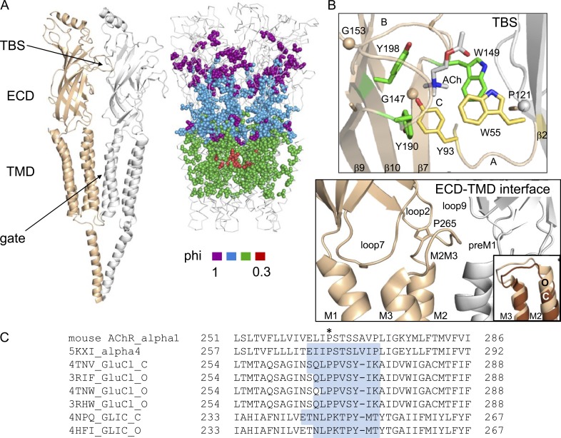
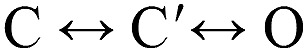
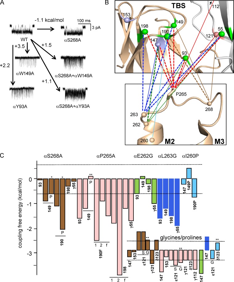
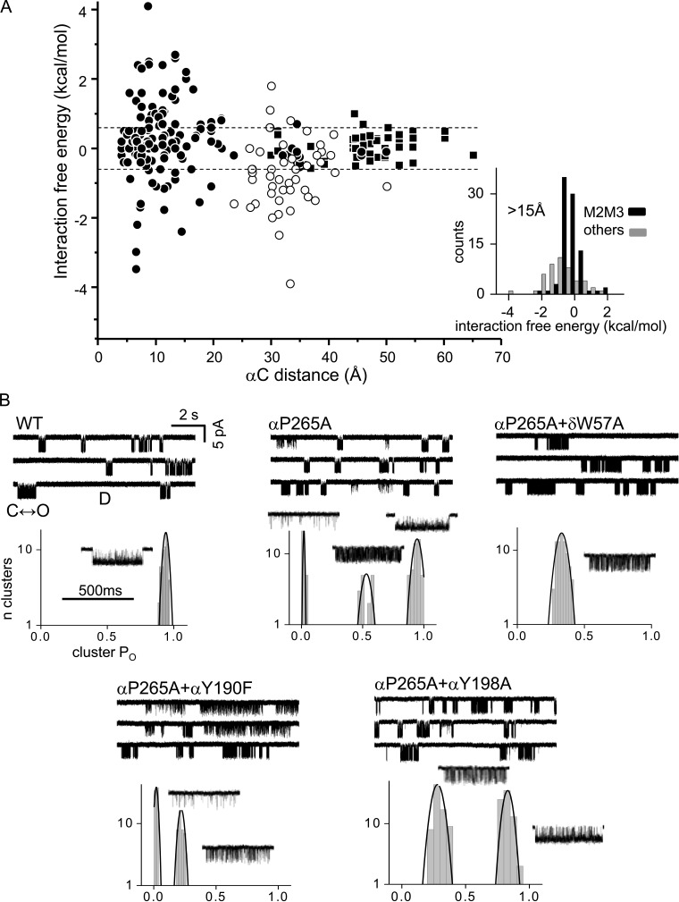
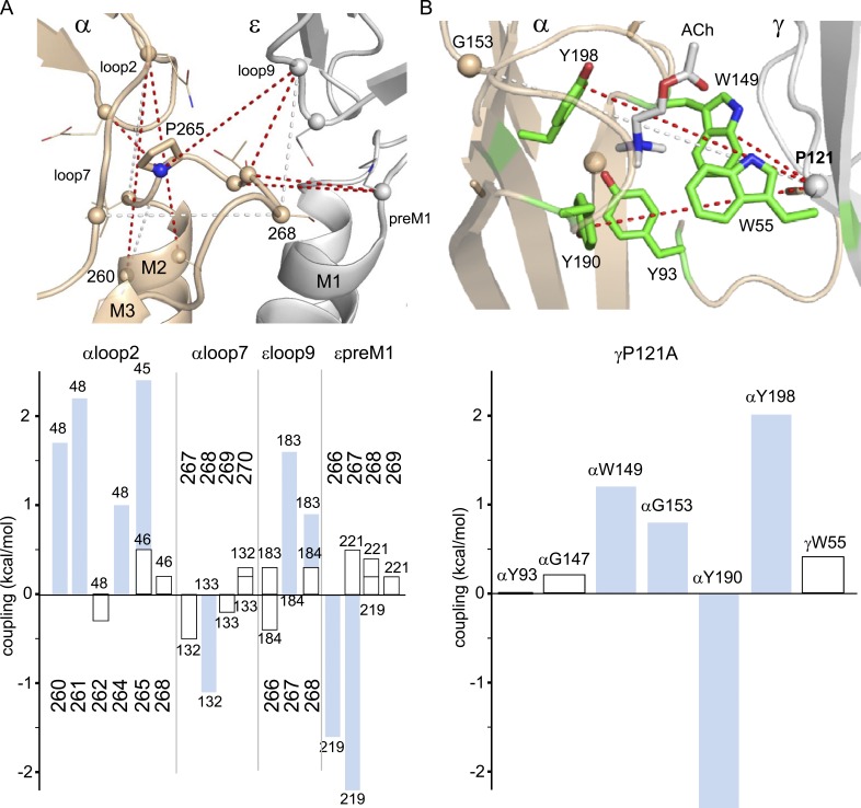
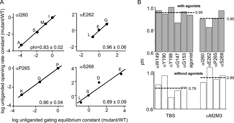
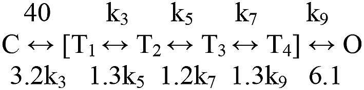
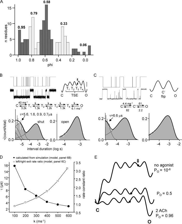
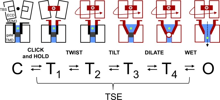
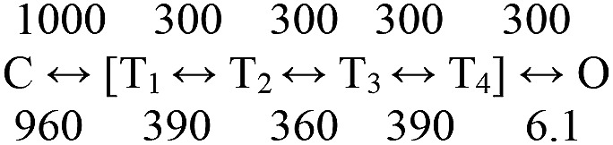
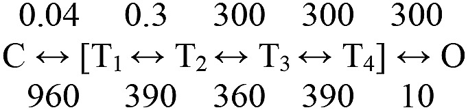
Nicotinic acetylcholine receptors are allosteric proteins that generate membrane currents by isomerizing ("gating") between resting and active conformations under the influence of neurotransmitters. Here, to explore the mechanisms that link the transmitter-binding sites (TBSs) with the distant gate, we use mutant cycle analyses to measure coupling between residue pairs, phi value analyses to sequence domain rearrangements, and current simulations to reproduce a microsecond shut component ("flip") apparent in single-channel recordings. Significant interactions between amino acids separated by >15 Å are rare; an exception is between the αM2-M3 linkers and the TBSs that are ∼30 Å apart. Linker residues also make significant, local interactions within and between subunits. Phi value analyses indicate that without agonists, the linker is the first region in the protein to reach the gating transition state. Together, the phi pattern and flip component suggest that a complete, resting↔active allosteric transition involves passage through four brief intermediate states, with brief shut events arising from sojourns in all or a subset. We derive energy landscapes for gating with and without agonists, and propose a structure-based model in which resting→active starts with spontaneous rearrangements of the M2-M3 linkers and TBSs. These conformational changes stabilize a twisted extracellular domain to promote transmembrane helix tilting, gate dilation, and the formation of a "bubble" that collapses to initiate ion conduction. The energy landscapes suggest that twisting is the most energetically unfavorable step in the resting→active conformational change and that the rate-limiting step in the reverse process is bubble formation.


Figures

Figure 1.
Figure 1.
Structures. (A, left) Torpedo AChR (PDB accession code 2BG9): α subunit, tan; γ subunit, white. The TBS is ∼50 Å from the gate. (right) AChR α subunit diliganded gating phi values mapped onto Caenorhabditis elegans GluCl (PDB accession code 3RIF); colors assigned by statistical criteria (Fig. 6 A; Purohit et al., 2013). Amino acids at the TBS and in αM2M3 have phi ∼1 (purple) and are separated by a domain of phi ∼0.8 residues. For clarity, some amino acids were removed at the ECD–TMD interface and M3. (B, top) Ligand-binding site of the Lymnaea stagnalis ACh-binding protein (PDB accession code 3WIP; AChR numbers). In adult AChRs, affinity is mainly determined by αY190, αY198, and αW149 (green), but in fetal AChRs, αY93 and γW55 also contribute (yellow). (bottom) ECD–TMD interface of human α4β2 AChRs (PDB accession code 5KXI). (inset) In GluCl, the M2 helix is displaced upward relative to M1 in O versus C (PDB accession codes 3RIF and 4TNV). (C) M2M3 sequence alignments. *, αP265 in mouse muscle AChRs. Highlighted residues are loop.
(Scheme 1)
(Scheme 1)
(Scheme 2)
(Scheme 2)
Figure 2.
Figure 2.
αM2M3–TBS coupling. (A) Examples of MCA. Single-channel currents are C↔O clusters (see Fig. 3 B); open is down. Arrows, gating free energy change caused by the mutation or mutations. αS268 is in αM2M3, and αW149/αY93 are at the TBS; αY93 shows no interaction (pair = sum), but αW149 is coupled significantly (sum > pair by 0.9 kcal/mol). (B) Map of αM2M3–TBS coupling. Connecting lines mark interacting amino acids: thick-dashed, strong (>1.2 kcal/mol); and thin-solid, significant (0.6–1.2 kcal/mol). TBS structure is AChBP (PDB accession code 3WIP), and M2M3 structure is α4β2 AChR (PDB accession code 5KXI); spheres are αC (TBS: green, aromatic; pink, proline; blue, glycine). (C) MCA coupling values. Dashed lines are at ±0.6 kcal/mol. Main, TBS aromatics; inset, TBS prolines and glycines. TBS mutations were to Ala except where indicated; the M2M3 mutations are shown except for *, Pro and **, Gly. All pairs are adult-type AChRs except for αY190F and αY198A (f, for fetal; 1 and 2 indicate modes; see Fig. 3 B).
Figure 3.
Figure 3.
Coupling summary and modal activity. (A) Interaction energy versus separation. αC distance is that between α-carbons of the mutations using GluCl as the template structure. Circles, MCA coupling free energy (open, αM2M3–TBS); squares, TMD–TBS interaction energies estimated from gating constants (see Materials and methods). Lines mark ±0.6 kcal/mol. (inset) Histograms of coupling energies for separations >15 Å (mean ± SD [n]): αM2M3–TBS, −0.68 ± 1.02 (47); all others, 0.09 ± 0.56 (87). (B) αP265A induces kinetic modes. Clusters reflect C↔O gating (O is down) and long silent periods are D(esensitized). WT clusters are homogeneous (PO = 0.88 ± 0.02; mean ± SD), αP265A are modal (0.02 ± 0.01, 0.51 ± 0.07, and 0.94 ± 0.03), and αP265A+δW57A are homogeneous (0.34 ± 0.03). δW57A eliminates modes, but αY190F and αY198A do not.
Figure 4.
Figure 4.
Short-range coupling. (A) Short-range αM2M3 coupling at the ECD–TMD interface (PDB accession code 5KXI; mouse α subunit muscle AChR numbering). Lines: red, strong; white, significant. αP265 interacts strongly with αloop2 (Q48 and E45), αM2 (I260), and εloop9 (G183). (bottom) MCA values; blue bars are significant (≥0.6 kcal/mol). (B) Interactions across the fetal αγ TBS interface (PDB accession code 3WIP).
Figure 5.
Figure 5.
Phi analysis of the TBS and αM2M3. (A) Unliganded gating REFERs for αM2M3 amino acids. The slope (phi) is given in each plot. (B) Comparison of phi values with versus without agonists (dashed lines, mean). For TBS residues, phi is higher with versus without agonists, but for αM2M3 residues, it is the same.
(Scheme 3)
(Scheme 3)
Figure 6.
Figure 6.
Combining phi and flip. (A) Phi values in diliganded AChR gating (Purohit et al., 2013). There are five populations, the first four of which correspond in decreasing order to purple, blue, green, and red in Fig. 1 A. (B) Simulations based on a model with four intermediate states (center; k = 300 ms−1; see supplemental text). (middle) The TSE are states T1–4 (all shut); each state is associated with a cartoon structure is in Fig. 7. (top left) Currents and perfect idealization. (top right) Energy landscape corresponding to the model (calibration, 1 kcal/mol; filled circle/‡, committor). (bottom) Histograms; sojourns in the TSE generate five shut components (gray lines), four of which are short-lived (time constants given); open sojourns are described by a single exponential. In standard patch recordings (∼25-µs resolution), the time constant of the open (long-shut) component is the inverse of the C↔O closing (opening) rate constant. (C) Fitting the simulations of panel B by C↔C′↔O after low-pass filtering to 30 kHz. There is only a single, brief component (flip/primed) apparent in the shut interval duration histogram. (D) Simulations using different values for k. Left y axis is the slowest shut interval TSE component (filled circles) calculated from the model, and right y axis (open squares) is the fitted backward/forward exit rate constant ratio from C′ using Scheme 2 (panel C). (E) Energy landscapes. (middle) The model in panel B with k = 300 ms−1; (top) the first two steps of unliganded opening are steeply uphill; (bottom) opening with two bound ACh molecules starts rapidly. In all conditions, the second step of the opening process (ECD twisting) is relatively the most difficult energetically, and the closing process is rate limited by gate bubble formation.
Figure 7.
Figure 7.
Structure-based model of AChR gating. Eight components undergo local off↔on transitions (black↔red): five are protein (left labels) plus the agonist (black/red filled circle), ions (green filled circle), and water (blue). Only the end state C (all components off) and O (all on) structures are stable. The gate region can be a hydrophobic cluster (gray square), a bubble (red open circle), or water. The four, short-lived intermediate states of the TSE (T1–4) have a mixture of on/off components. The five steps in the opening process are M2M3–TBS click-and-hold, ECD twist, TMD tilt, gate dilate, and gate wetting (bubble collapse). The energy landscapes (Fig. 6 E and Fig. S2) calculated from phi and flip suggest that ECD twisting is the most energetically unfavorable step in the opening process. Channel closing is the reverse of opening and starts with (and is rate limited by) the spontaneous reformation of a gate bubble. Animations of this scheme at different time scales are shown in Video 1.
(Scheme 4)
(Scheme 4)
(Scheme 5)
(Scheme 5)

References

    1. Althoff T., Hibbs R.E., Banerjee S., and Gouaux E.. 2014. X-ray structures of GluCl in apo states reveal a gating mechanism of Cys-loop receptors. Nature. 512:333–337. 10.1038/nature13669 - DOI - PMC - PubMed
    1. Auerbach A. 2005. Gating of acetylcholine receptor channels: Brownian motion across a broad transition state. Proc. Natl. Acad. Sci. USA. 102:1408–1412. 10.1073/pnas.0406787102 - DOI - PMC - PubMed
    1. Auerbach A. 2010. The gating isomerization of neuromuscular acetylcholine receptors. J. Physiol. 588:573–586. 10.1113/jphysiol.2009.182774 - DOI - PMC - PubMed
    1. Auerbach A. 2012. Thinking in cycles: MWC is a good model for acetylcholine receptor-channels. J. Physiol. 590:93–98. 10.1113/jphysiol.2011.214684 - DOI - PMC - PubMed
    1. Bafna P.A., Purohit P.G., and Auerbach A.. 2008. Gating at the mouth of the acetylcholine receptor channel: energetic consequences of mutations in the alphaM2-cap. PLoS One. 3:e2515 10.1371/journal.pone.0002515 - DOI - PMC - PubMed

Substances

LinkOut - more resources