Review
. 2016 Nov 24:10:2345-2361.
doi: 10.2147/OPTH.S94866. eCollection 2016.

Vogt-Koyanagi-Harada syndrome - current perspectives

Affiliations
Review

Vogt-Koyanagi-Harada syndrome - current perspectives

Abeir Baltmr et al. Clin Ophthalmol. .

Abstract

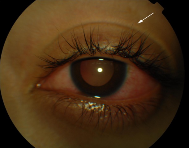
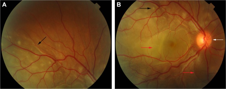
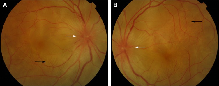
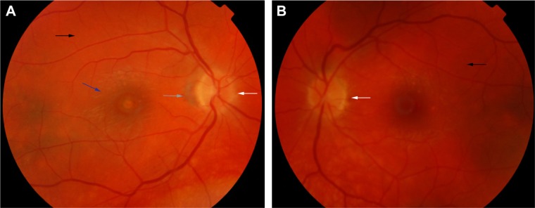
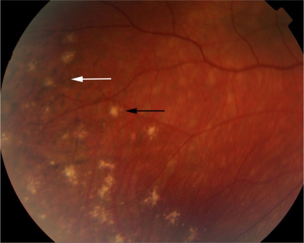
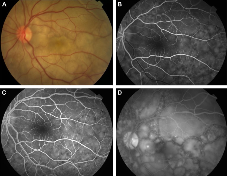
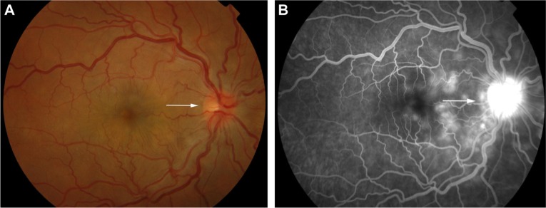
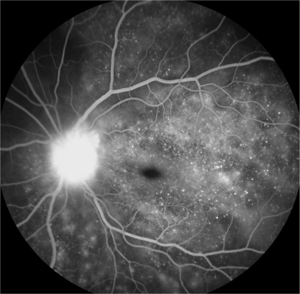
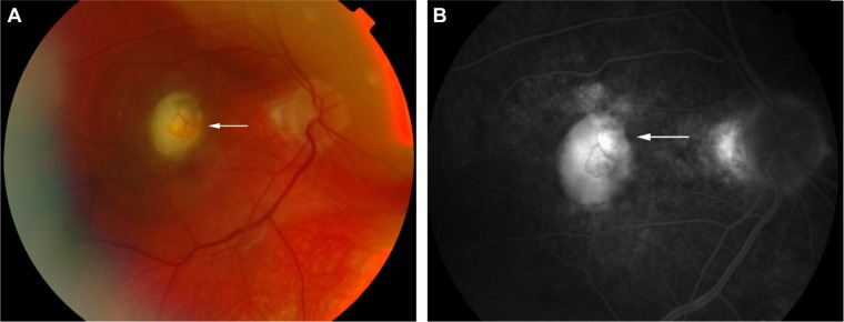
Vogt-Koyanagi-Harada syndrome is a cause of noninfectious panuveitis, leading to significant vision loss in many patients. It is an autoimmune disease occurring in genetically susceptible individuals and clinically presents as bilateral panuveitis with serous retinal detachments and hyperemic, swollen optic discs, which are associated with neurological and auditory manifestations. Early diagnosis and prompt and adequate treatment with immunosuppressive agents (corticosteroids and other immunosuppressive drugs) may halt disease progression and prevent recurrences and vision loss. This review summarizes the current knowledge on the variable clinical aspects of this disease, highlighting diagnostic and treatment strategies.

Keywords: corticosteroid; multifocal choroiditis; panuveitis; serous retinal detachment; starry sky; sunset glow fundus.


Conflict of interest statement

The authors report no conflicts of interest in this work.

Figures

Figure 1
Figure 1
Color photograph of a patient in the chronic recurrent phase of the VKHD showing acute anterior uveitis and mild poliosis (white arrow). Abbreviation: VKHD, Vogt–Koyanagi–Harada disease.
Figure 2
Figure 2
Color retinal photographs of a patient during the acute phase of VKHD. Notes: (A) Multifocal choroiditis lesions of variable size representing along the superior arcade (black arrow). (B) The posterior pole depicting a hyperemic optic disc (white arrow), multifocal choroiditis (black arrow), and multiple areas of SRDs (red arrows). Abbreviations: VKHD, Vogt–Koyanagi–Harada disease; SRDs, serous retinal detachments.
Figure 3
Figure 3
Color retinal photographs of right (A) and left eye (B) showing swollen and hyperemic optic discs (white arrows), with choroidal folds disc (black arrows), in the acute phase of VKHD. Abbreviation: VKHD, Vogt–Koyanagi–Harada disease.
Figure 4
Figure 4
Color retinal photographs of right (A) and left eye (B) during the convalescent phase of VKHD, showing sunset glow fundus with pale optic discs (white arrows) and bright-orange choroids (black arrows). Note the peripapillary atrophy (gray arrow) and macular scaring (blue arrow) in (A). Abbreviation: VKHD, Vogt–Koyanagi–Harada disease.
Figure 5
Figure 5
Color retinal photograph displaying sunset glow fundus with a numerous small choroidal depigmented atrophic lesions (black arrow) and hyperpigmented lesions (white arrow) in the retinal periphery during the convalescent phase of VKHD. Abbreviation: VKHD, Vogt–Koyanagi–Harada disease.
Figure 6
Figure 6
Color retinal and FFA photographs of a patient in the acute phase of VKHD showing (A) hyperemic optic disc with deep yellow lesions of variable size and multiple SRD, (B) early FFA showing delayed choroidal filling, (C) mid phase of FFA showing pinpoint hyperfluorescent leakage at the RPE level (D) with pooling of the dye in the subretinal space in the late phase of the angiogram. Abbreviations: FFA, fundus fluorescein angiography; VKHD, Vogt–Koyanagi–Harada disease; SRD, serous retinal detachment; RPE, retinal pigment epithelium.
Figure 7
Figure 7
Color retinal photograph of the right eye showing swollen and hyperemic optic discs (A) and FFA showing hot disc (B) (white arrows), in the acute phase of VKHD. Abbreviations: FFA, fundus fluorescein angiography; VKHD, Vogt–Koyanagi–Harada disease.
Figure 8
Figure 8
FFA photograph of a patient in the acute phase of VKHD showing optic disc leakage and numerous hyperfluorescent pinpoint foci of leakage at the level of RPE leading to the classic “starry sky” appearance. Abbreviations: FFA, fundus fluorescein angiography; VKHD, Vogt–Koyanagi–Harada disease; RPE, retinal pigment epithelium.
Figure 9
Figure 9
Color retinal (A) and FFA (B) photographs of a VKHD patient with macular CNV (white arrows). Abbreviations: FFA, fundus fluorescein angiography; VKHD, Vogt–Koyanagi–Harada disease; CNV, choroidal neovascularization.
Figure 10
Figure 10
ICG photographs of the right eye of a patient in the acute phase of VKHD showing (A) patchy hypofluorescence during the early angiographic phase, (B) large choroidal stromal vessel hyperfluorescence with fuzzy choroidal vessels in the early phase, (C) hypofluorescent dark dots during the intermediate phase of angiography, and (D) diffuse choroidal hyperfluorescence in the late phase. Abbreviations: ICG, indocyanine green angiography; VKHD, Vogt–Koyanagi–Harada disease.
Figure 11
Figure 11
Short wavelength light (blue) FAF photographs of eyes of a patient with chronic VKHD. Notes: (A) Right eye showing areas of hypoautofluorescence (black arrow) corresponding to RPE atrophy and loss. (B) Left eye showing areas of hyperautofluorescence (white arrow) corresponding to RPE proliferation. Abbreviations: FAF, fundus autofluorescence; VKHD, Vogt–Koyanagi–Harada disease; RPE, retinal pigment epithelium.
Figure 12
Figure 12
OCT scan of the macula of a patient in the acute phase of VKHD before and after starting corticosteroid treatment. Notes: (A) SRD (white arrow) and a mild fluctuation in the ILM (red arrow). (B) Resolution of SRD with treatment (white arrow). Abbreviations: OCT, optical coherence tomography; VKHD, Vogt–Koyanagi–Harada disease; SRD, serous retinal detachment; ILM, internal limiting membrane.

References

    1. Moorthy RS, Inomata H, Rao NA. Vogt–Koyanagi–Harada syndrome. Surv Ophthalmol. 1995;39(4):265–292. - PubMed
    1. Read RW, Rechodouni A, Butani N, et al. Complications and prognostic factors in Vogt–Koyanagi–Harada disease. Am J Ophthalmol. 2001;131(5):599–606. - PubMed
    1. Norose K, Yano A. Melanoma specific Th1 cytotoxic T lymphocyte lines in Vogt–Koyanagi–Harada disease. Br J Ophthalmol. 1996;80(11):1002–1008. - PMC - PubMed
    1. Yamaki K, Gocho K, Hayakawa K, Kondo I, Sakuragi S. Tyrosinase family proteins are antigens specific to Vogt–Koyanagi–Harada disease. J Immunol. 2000;165(12):7323–7329. - PubMed
    1. Sugita S, Takase H, Taguchi C, et al. Ocular infiltrating CD4+ T cells from patients with Vogt–Koyanagi–Harada disease recognize human melanocyte antigens. Invest Ophthalmol Vis Sci. 2006;47(6):2547–2554. - PubMed

LinkOut - more resources