. 2016 Nov 25:7:452.
doi: 10.3389/fphar.2016.00452. eCollection 2016.

Proton Pump Inhibitors Display Antitumor Effects in Barrett's Adenocarcinoma Cells

Affiliations

Proton Pump Inhibitors Display Antitumor Effects in Barrett's Adenocarcinoma Cells

Eduardo Chueca et al. Front Pharmacol. .

Abstract

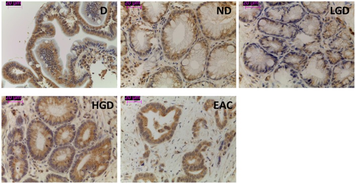
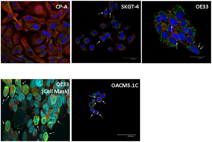
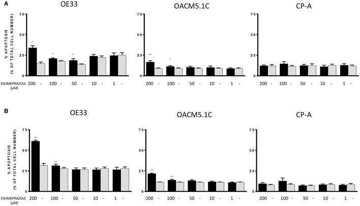
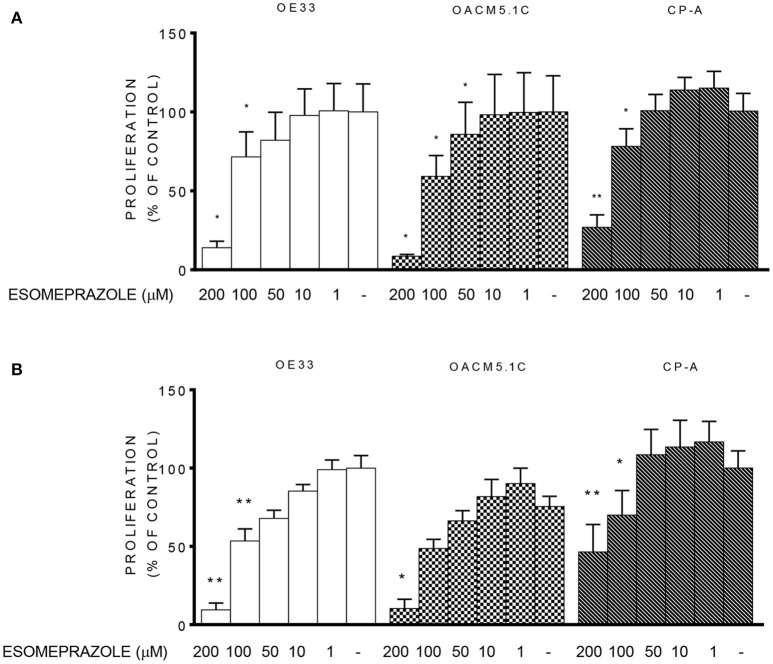
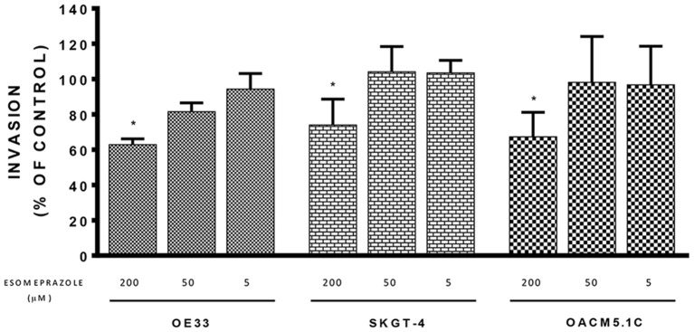
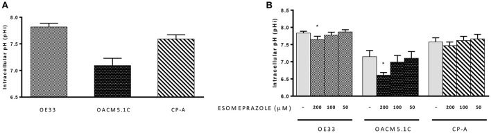
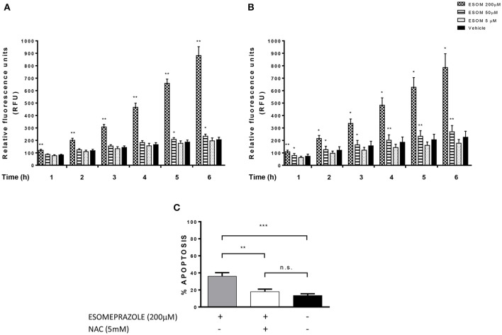
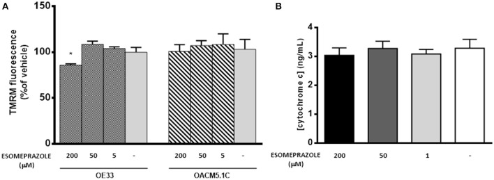
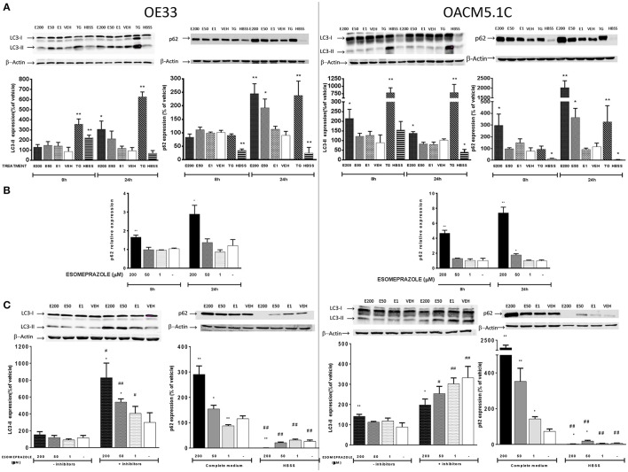
Recent evidence has reported that proton pump inhibitors (PPIs) can exert antineoplastic effects through the disruption of pH homeostasis by inhibiting vacuolar ATPase (H+-VATPase), a proton pump overexpressed in several tumor cells, but this aspect has not been deeply investigated in EAC yet. In the present study, the expression of H+-VATPase was assessed through the metaplasia-dysplasia-adenocarcinoma sequence in Barrett's esophagus (BE) and the antineoplastic effects of PPIs and cellular mechanisms involved were evaluated in vitro. H+-VATPase expression was assessed by immunohistochemistry in paraffined-embedded samples or by immunofluorescence in cultured BE and EAC cell lines. Cells were treated with different concentrations of PPIs and parameters of citotoxicity, oxidative stress, and autophagy were evaluated. H+-VATPase expression was found in all biopsies and cell lines evaluated, showing differences in the location of the pump between the cell lines. Esomeprazole inhibited proliferation and cell invasion and induced apoptosis of EAC cells. Production of reactive oxygen species (ROS) seemed to be involved in the cytotoxic effects observed since the addition of N-acetylcysteine significantly reduced esomeprazole-induced apoptosis in EAC cells. Esomeprazole also reduced intracellular pH of tumor cells, whereas only disturbed the mitochondrial membrane potential in OE33 cells. Esomeprazole induced autophagy in both EAC cells, but also triggered a blockade in autophagic flux in the metastatic cell line. These data provide in vitro evidence supporting the potential use of PPIs as novel antineoplastic drugs for EAC and also shed some light on the mechanisms that trigger PPIs cytotoxic effects, which differ upon the cell line evaluated.

Keywords: Barrett's esophagus; esophageal adenocarcinoma; proton pump inhibitors; reactive oxygen species; vacuolar ATPase.


Figures

Figure 1
Figure 1
Representative images of V-ATPase expression in BE and EAC in biopsy samples. Immunohistochemical labeling of V-ATPase (brown staining) in paraffin-embedded biopsy samples corresponding to duodenum and the different stages of neoplastic progression in BE and EAC.
Figure 2
Figure 2
Representative images of V-ATPase expression in BE and EAC cell lines. Confocal microscopy showing V-ATPase (green dots) and plasma membrane (red/orange staining) in BE and EAC cell lines.
Figure 3
Figure 3
Effects of esomeprazole on apoptosis of EAC and BE cell lines. Apoptosis was evaluated in EAC and BE cell lines under physiological (A) or acidified (B) conditions. The bars represent the mean % of apoptosis in esomeprazole-treated cells with respect to control cells (DMSO only). All data are expressed as mean ± SEM of at least three independent experiments. Significant level *p < 0.05; **p < 0.01.
Figure 4
Figure 4
Effects of esomeprazole on cell proliferation. Cell proliferation in EAC and BE cell lines at physiological (A) or acidified (B) culture medium. The results are represented as the percentage of BrdU incorporation in esomeprazole-treated cells in comparison with untreated controls (DMSO only). All data are expressed as mean ± SEM of at least three independent experiments. Significant level *p < 0.05; **p < 0.01.
Figure 5
Figure 5
Effects of esomeprazole on cell invasion. The effects of esomeprazole on cell invasive properties of EAC cells were expressed as fluorescence values (RFUs) in esomeprazole-treated cells relative to untreated cells. All data are expressed as mean ± SEM of at least three independent experiments. Significant level *p < 0.05.
Figure 6
Figure 6
Intracellular pH. Basal pHi levels of OE33, OACM5.1C and CP-A cells (A). Effects of esomeprazole on pHi (B). Data are represented as the mean ± SEM of three independent experiments. Significant differences from the respective control values:*p < 0.05.
Figure 7
Figure 7
Esomeprazole and ROS production in EAC cells. ROS levels in esomeprazole-treated OE33 (A) and OACM5.1C (B) cells and effects of the antioxidant NAC on esomeprazole-induced apoptosis in OE33 cells (C). All data are expressed as the mean ± SEM of at least three independent experiments. Significant differences: *p < 0.05; **p < 0.01; ***p < 0.001.
Figure 8
Figure 8
Effects of esomeprazole on mitochondrial membrane potential (A) and cytochrome C release from mitochondria to cytosol (B). Data showed in (A) are represented as % of TMRM fluorescence intensity in esomeprazole-treated cells with respect to vehicle. Data showed in (B) are expressed as mean ± SEM of cytosolic concentration of cytochrome c in esomeprazole-treated OE33 cells. All experiments were repeated at least three times. Significant differences from the respective control values: *p < 0.05.
Figure 9
Figure 9
Effects on autophagy of EAC cells. OE33 and OACM5.1C cells were used. Autophagy markers LC3 and p62 were evaluated by WB after 8 and 24 h incubation with esomeprazole (A). p62 relative expression (B) was evaluated by RT-PCR after treatment with esomeprazole for 8 and 24 h and the results are expressed as the level of expression of p62 with respect to cells treated with vehicle alone. β-actin was used as a housekeeper control. The effects of esomeprazole on autophagic flux (C) was evaluated by WB after incubating cells in complete media in the presence or absence of lysosomal inhibitors pepstatin A and E-64d, and in complete media or HBSS for 24 h. WB representative gels are lined up their respective results and all data are represented as the % of expression of autophagy markers LC3-II or p62 with respect to values obtained in cells treated with vehicle alone. All data are represented as mean ± SEM of three independent experiments. Significant differences from the respective control values: *p < 0.05; **p < 0.01. Significant differences from the respective treatment in complete medium or medium without inhibitors: #p < 0.05; ##p < 0.01.

References

    1. Azzarito T., Venturi G., Cesolini A., Fais S. (2015). Lansoprazole induces sensitivity to suboptimal doses of paclitaxel in human melanoma. Cancer Lett. 356, 697–703. 10.1016/j.canlet.2014.10.017 - DOI - PubMed
    1. Barth S., Glick D., Macleod K. F. (2010). Autophagy: assays and artifacts. J. Pathol. 221, 117–124. 10.1002/path.2694 - DOI - PMC - PubMed
    1. Chow S. H., Hedley D. (1997). Flow cytometric measurement of intracellular pH. Curr. Protoc. Cytom. Chapter 9:Unit 9.3. 10.1002/0471142956.cy0903s14 - DOI - PubMed
    1. Chung C., Mader C. C., Schmitz J. C., Atladottir J., Fitchev P., Cornwell M. L., et al. . (2011). The vacuolar-ATPase modulates matrix metalloproteinase isoforms in human pancreatic cancer. Lab. Invest. 91, 732–743. 10.1038/labinvest.2011.8 - DOI - PMC - PubMed
    1. Cregan S. P., Dawson V. L., Slack R. S. (2004). Role of AIF in caspase-dependent and caspase-independent cell death. Oncogene 23, 2785–2796. 10.1038/sj.onc.1207517 - DOI - PubMed

LinkOut - more resources