. 2016 Dec 1;2(2):e000349.
doi: 10.1136/rmdopen-2016-000349. eCollection 2016.

Spontaneous destructive periodontitis and skeletal bone damage in transgenic mice carrying a human shared epitope-coding HLA-DRB1 allele

Affiliations

Spontaneous destructive periodontitis and skeletal bone damage in transgenic mice carrying a human shared epitope-coding HLA-DRB1 allele

Prashasnika Gehlot et al. RMD Open. .

Abstract

Objective: Shared epitope (SE)-coding DRB1 alleles are associated with bone erosion in several diseases, including rheumatoid arthritis (RA) and periodontal disease (PD), but the underlying mechanism is unknown. We have recently identified the SE as an osteoclast-activating ligand. To better understand the biological effects of the SE in vivo, here we sought to determine whether it can facilitate spontaneous bone damage in naïve mice.

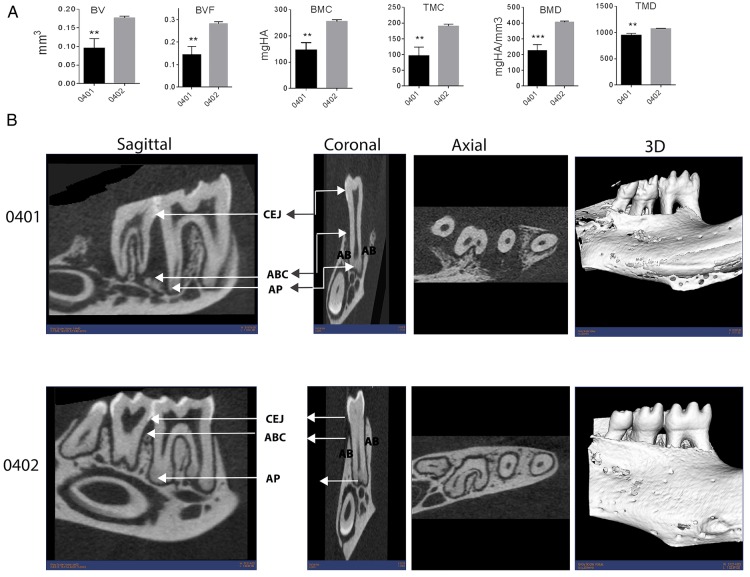
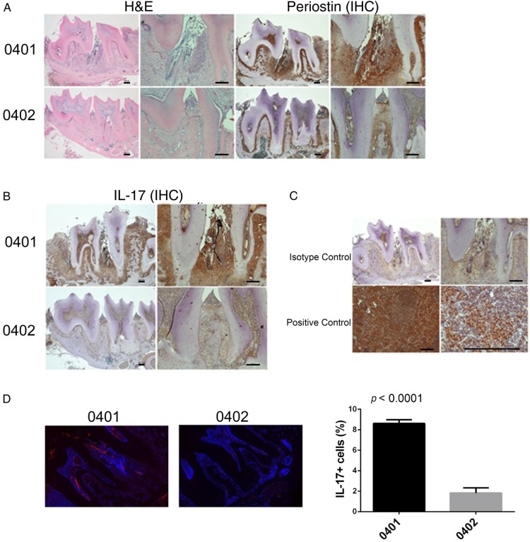
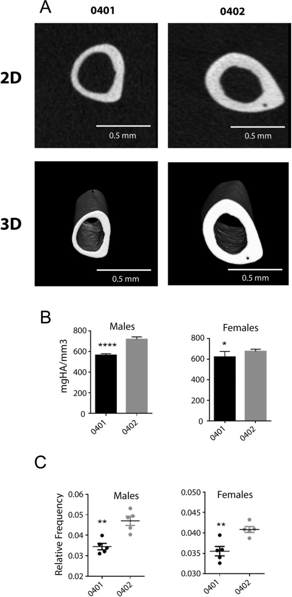
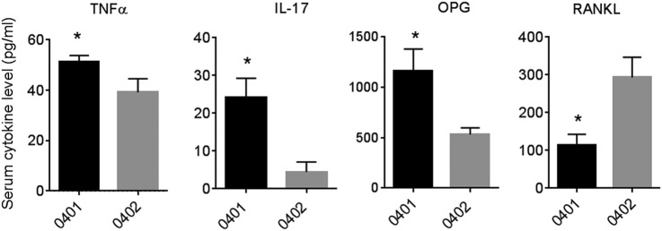
Methods: 3-month old naïve transgenic mice that carry the human SE-coding allele DRB1*04:01, or a SE-negative allele DRB1*04:02 were studied. Bone tissues were analysed by micro-CT, and the tooth-supporting tissues were studied by histology, immunohistochemistry and immunofluorescence. Serum biomarkers were determined by ELISA.

Results: Transgenic mice expressing the SE-coding DRB1*04:01 allele, but not mice carrying the SE-negative allele DRB1*04:02, showed spontaneous PD associated with interleukin (IL)-17 overabundance and periostin disruption. Mandibular bone volumetric and mineralisation parameters were significantly lower in SE-positive mice, and alveolar bone resorption was significantly increased in these mice. SE-positive mice also had more slender tibiae, and their marrow, cortical and total areas were lower than those of SE-negative mice. Additionally, significantly increased serum IL-17, tumour necrosis factor-α and osteoprotegrin levels were found in SE-positive mice, while their receptor activator of nuclear factor κ-B ligand levels were significantly lower.

Conclusions: A human SE-coding allele increases the propensity to spontaneous bone-destructive periodontal inflammation and skeletal bone damage in transgenic mice. These findings provide new insights into the previously documented but poorly understood association of the SE with accelerated bone erosion in RA and several other human diseases.

Keywords: Bone Mineral Density; Gene Polymorphism; Rheumatoid Arthritis.


Conflict of interest statement

Conflicts of Interest: None declared.

Figures

Figure 1
Figure 1
Periodontal micro-CT analyses. (A) 3D micro-CT-based quantification of mandibular bone parameters in SE-positive DRB1*04:01 (black bars) and SE-negative DRB1*04:02 (gray bars) Tg mice. Values represent mean and SEM (n=5 per group). (B) Sagittal, coronal and axial 2D (left 3 columns) and 3D (right column) micro-CT images of mandibles from representative SE-positive DRB1*04:01 (upper panel) and SE-negative DRB1*04:02 (lower panel) Tg male mice. 2D, two-dimensional; 3D, three-dimensional; ABC, alveolar bone crest; AP, anteroposterior; BV, bone volume; BMC, bone mineral content; BMD, bone mineral density; CEJ, cemento enemal junction; BVF, bone volume fraction; SE, shared epitope; TMC, tissue mineral content; TMD, tissue mineral density.
Figure 2
Figure 2
Spontaneous PD in DRB1*04:01 mice. (A) Representative mandibular tissue sections stained with H&E (left) show intense inflammatory infiltrate (left). IHC staining for periostin (right) show disruption of the periodontal ligament in SE-positive, DRB1*04:01 Tg mice (top panel). SE-negative DRB1*04:02 Tg mice (bottom panel) show normal histology. (B) IHC staining for IL-17A showing markedly increased abundance of the cytokine in mandibular periodontal tissue of SE-positive, DRB1*04:01 Tg mice (top panel), compared with SE-negative, DRB1*04:02 Tg mice (bottom panel). (C) Isotype-matched antibody control shows negative IHC staining (top panel). A positive control tissue (draining lymph node—10× and 40×) shows the expected abundance of IL-17. In H&E and IHC, images of low (4×, left) and high (10×, right) magnification are shown. Horizontal bars represent 100 µm. (D) Identification of IL-17-positive cells in periodontal tissues by immunofluorescence. Red fluorescence represents IL-17; blue fluorescence (DAPI) identifies nuclei. Boxed images show higher magnification images of regions identified by white arrows. Bar graph on the right depicts mean and SEM of IL-17-positive cells in SE-positive, DRB1*04:01 Tg mice (black) and SE-negative, DRB1*04:02 Tg mice (gray), as quantified by a Biotek Cytation5 instrument. DAPI, 4', 6-diamidino-2-phenylindole; IHC, immunohistochemistry; IL, interleukin; SE, shared epitope; PD, periodontal disease.
Figure 3
Figure 3
Tibial micro-CT analysis. (A) Representative 2D (upper panel) and 3D (lower panel) tibiae micro-CT images of SE-positive DRB1*04:01 (left column) and SE-negative DRB1*04:02 (right column) mice. (B) Tibial mineral density data of SE-positive DRB1*04:01 (black bars) and SE-negative DRB1*04:02 (gray bars) mice. (C) Male (left) and female (right) robustness data of the SE-positive DRB1*04:01 (black dots) and SE-negative DRB1*04:02 (gray dots) mouse groups. Values in (B) and (C) represent mean and SEM (n=5 per group). 2D, two-dimensional; 3D, three-dimensional; SE, shared epitope.
Figure 4
Figure 4
Serological markers. Serum concentrations of TNF-α, IL-17, RANKL and OPG in SE-positive DRB1*04:01 (black bars) and SE-negative DRB1*04:02 (gray bars) mice. Values represent mean and SEM (n=5 per group). IL, interleukin; OPG, osteoprotegrin; RANKL, receptor activator of nuclear factor κ-B ligand; SE, shared epitope; TNF, tumour necrosis factor.

References

    1. Mercado FB, Marshall RI, Klestov AC et al. . Relationship between rheumatoid arthritis and periodontitis. J Periodontol 2001;72:779–87. 10.1902/jop.2001.72.6.779 - DOI - PubMed
    1. de Pablo P, Chapple IL, Buckley CD et al. . Periodontitis in systemic rheumatic diseases. Nat Rev Rheumatol 2009;5:218–24. 10.1038/nrrheum.2009.28 - DOI - PubMed
    1. da Silva AP, Bissada NF. “Arthritis and Periodontitis: an association debated for over two centuries”. Curr Rheumatol Rev Published Online First: 26 Oct 2015. - PubMed
    1. Miossec P, Korn T, Kuchroo VK. Interleukin-17 and type 17 helper T cells. N Engl J Med 2009;361:888–98. 10.1056/NEJMra0707449 - DOI - PubMed
    1. Gaffen SL, Hajishengallis G. A new inflammatory cytokine on the block: re-thinking periodontal disease and the Th1/Th2 paradigm in the context of Th17 cells and IL-17. J Dent Res 2008;87:817–28. 10.1177/154405910808700908 - DOI - PMC - PubMed

LinkOut - more resources