

Modeling Membrane Protein-Ligand Binding Interactions: The Human Purinergic Platelet Receptor
- PMID: 27934233
- PMCID: PMC5460638
- DOI: 10.1021/acs.jpcb.6b09535
Modeling Membrane Protein-Ligand Binding Interactions: The Human Purinergic Platelet Receptor
Abstract
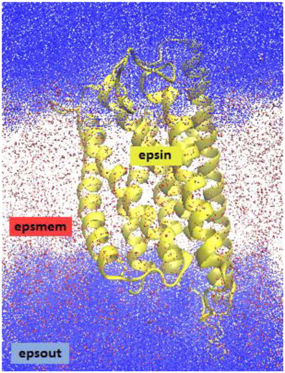
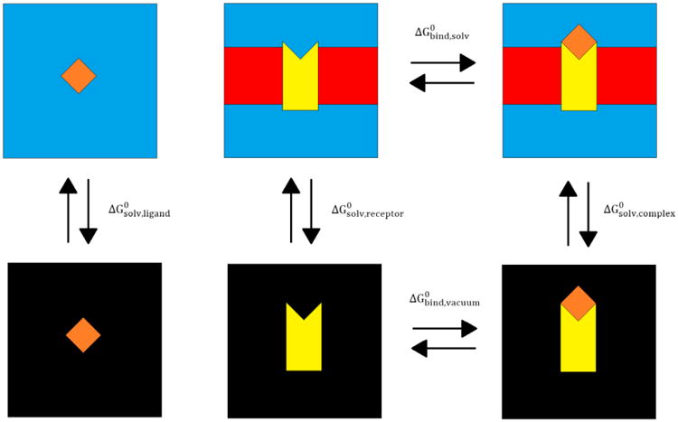
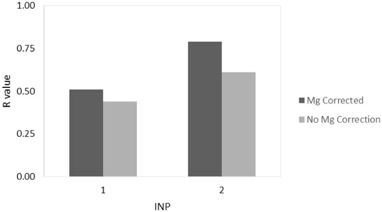
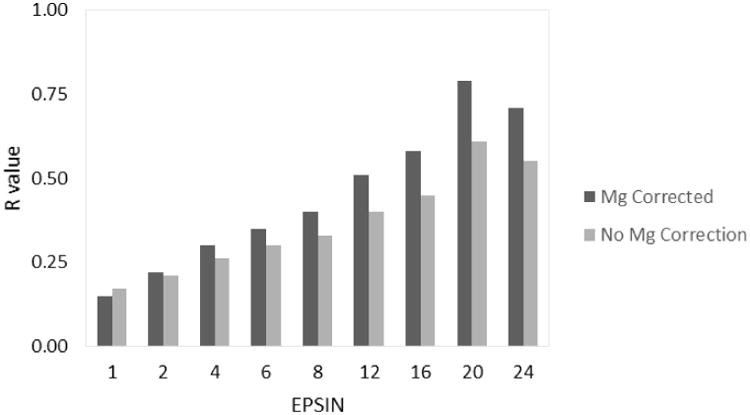
Membrane proteins, due to their roles as cell receptors and signaling mediators, make prime candidates for drug targets. The computational analysis of protein-ligand binding affinities has been widely employed as a tool in rational drug design efforts. Although efficient implicit solvent-based methods for modeling globular protein-ligand binding have been around for many years, the extension of such methods to membrane protein-ligand binding is still in its infancy. In this study, we extended the widely used Amber/MMPBSA method to model membrane protein-ligand systems, and we used it to analyze protein-ligand binding for the human purinergic platelet receptor (P2Y12R), a prominent drug target in the inhibition of platelet aggregation for the prevention of myocardial infarction and stroke. The binding affinities, computed by the Amber/MMPBSA method using standard parameters, correlate well with experiment. A detailed investigation of these parameters was conducted to assess their impact on the accuracy of the method. These analyses show the importance of properly treating the nonpolar solvation interactions and the electrostatic polarization in the binding of nucleotide agonists and non-nucleotide antagonists to P2Y12R. On the basis of the crystal structures and the experimental conditions in the binding assay, we further hypothesized that the nucleotide agonists lose their bound magnesium ion upon binding to P2Y12R, and our computational study supports this hypothesis. Ultimately, this work illustrates the value of computational analysis in the interpretation of experimental binding reactions.
Figures
Similar articles
-
Agonist-bound structure of the human P2Y12 receptor.Nature. 2014 May 1;509(7498):119-22. doi: 10.1038/nature13288. Nature. 2014. PMID: 24784220 Free PMC article.
-
Modeling ligand recognition at the P2Y12 receptor in light of X-ray structural information.J Comput Aided Mol Des. 2015 Aug;29(8):737-56. doi: 10.1007/s10822-015-9858-z. Epub 2015 Jul 21. J Comput Aided Mol Des. 2015. PMID: 26194851 Free PMC article.
-
Structure of the human P2Y12 receptor in complex with an antithrombotic drug.Nature. 2014 May 1;509(7498):115-8. doi: 10.1038/nature13083. Epub 2014 Mar 23. Nature. 2014. PMID: 24670650 Free PMC article.
-
P2Y12 receptors: structure and function.J Thromb Haemost. 2015 Jun;13 Suppl 1:S10-6. doi: 10.1111/jth.12952. J Thromb Haemost. 2015. PMID: 26149010 Review.
-
Clopidogrel: interactions with the P2Y12 receptor and clinical relevance.Hematology. 2003 Dec;8(6):359-65. doi: 10.1080/10245330310001621260. Hematology. 2003. PMID: 14668029 Review. No abstract available.
Cited by
-
Large scale peptide screening against main protease of SARS CoV-2.J Comput Chem. 2023 Mar 30;44(8):887-901. doi: 10.1002/jcc.27050. Epub 2022 Dec 7. J Comput Chem. 2023. PMID: 36478400 Free PMC article.
-
Targeting the receptor binding domain and heparan sulfate binding for antiviral drug development against SARS-CoV-2 variants.Sci Rep. 2024 Feb 2;14(1):2753. doi: 10.1038/s41598-024-53111-2. Sci Rep. 2024. PMID: 38307890 Free PMC article.
-
A Continuum Poisson-Boltzmann Model for Membrane Channel Proteins.J Chem Theory Comput. 2017 Jul 11;13(7):3398-3412. doi: 10.1021/acs.jctc.7b00382. Epub 2017 Jun 14. J Chem Theory Comput. 2017. PMID: 28564540 Free PMC article.
-
Mycobactin analogue interacting with siderophore efflux-pump protein: insights from molecular dynamics simulations and whole-cell assays.Front Antibiot. 2024 May 8;3:1362516. doi: 10.3389/frabi.2024.1362516. eCollection 2024. Front Antibiot. 2024. PMID: 39816270 Free PMC article.
-
Targeting ion channels with ultra-large library screening for hit discovery.Front Mol Neurosci. 2024 Jan 5;16:1336004. doi: 10.3389/fnmol.2023.1336004. eCollection 2023. Front Mol Neurosci. 2024. PMID: 38249296 Free PMC article. Review.
References
-
- Case DA, Betz RM, Botello-Smith W, Cerutti DS, T E Cheatham I, Darden TA, Duke RE, Giese TJ, Gohlke H, Goetz AW, et al. Amber 16. University of California, San Francisco; San Francisco, CA: 2016.
-
- Case DA, Betz RM, Botello-Smith W, Cerutti DS, T E Cheatham I, Darden TA, Duke RE, Giese TJ, Gohlke H, Goetz AW, et al. AmberTools 16. University of California, San Francisco; San Francsico, CA: 2016.
-
- Srinivasan J, Cheatham TE, Cieplak P, Kollman PA, Case DA. Continuum Solvent Studies of the Stability of DNA, RNA, and Phosphoramidate - DNA Helices. J Am Chem Soc. 1998;120:9401–9409.
-
- Kollman PA, Massova I, Reyes C, Kuhn B, Huo SH, Chong L, Lee M, Lee T, Duan Y, Wang W, et al. Calculating Structures and Free Energies of Complex Molecules: Combining Molecular Mechanics and Continuum Models. Acc Chem Res. 2000;33:889–897. - PubMed
-
- Gohlke H, Case DA. Converging Free Energy Estimates: MM-PB(GB)SA Studies on the Protein-Protein Complex Ras-Raf. J Comput Chem. 2004;25:238–250. - PubMed
Publication types
MeSH terms
Substances
Grants and funding
LinkOut - more resources
Full Text Sources
Other Literature Sources
