. 2016 Dec 2;354(6316):1144-1148.
doi: 10.1126/science.aah5715. Epub 2016 Nov 10.

Gliogenic LTP spreads widely in nociceptive pathways

Affiliations

Gliogenic LTP spreads widely in nociceptive pathways

M T Kronschläger et al. Science. .

Abstract

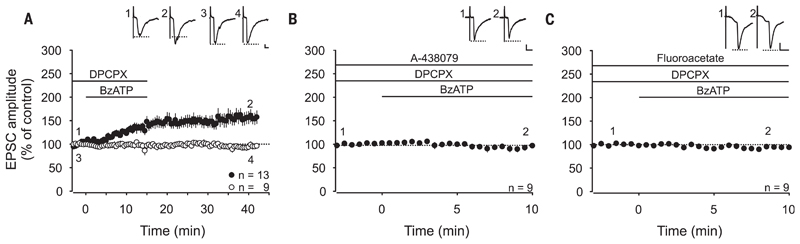
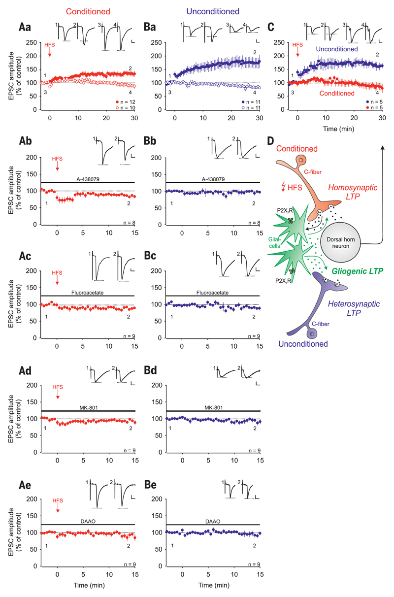
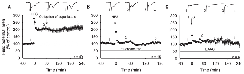
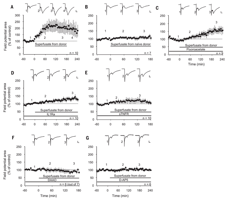
Learning and memory formation involve long-term potentiation (LTP) of synaptic strength. A fundamental feature of LTP induction in the brain is the need for coincident pre- and postsynaptic activity. This restricts LTP expression to activated synapses only (homosynaptic LTP) and leads to its input specificity. In the spinal cord, we discovered a fundamentally different form of LTP that is induced by glial cell activation and mediated by diffusible, extracellular messengers, including d-serine and tumor necrosis factor (TNF), and that travel long distances via the cerebrospinal fluid, thereby affecting susceptible synapses at remote sites. The properties of this gliogenic LTP resolve unexplained findings of memory traces in nociceptive pathways and may underlie forms of widespread pain hypersensitivity.


Conflict of interest statement

The authors declare no conflicts of interest. M.T.K., R.D.-S., and J.S. designed the research. M.T.K., R.D.-S., M.G., S.D.H., and H.L.T. generated and analyzed the data. M.T.K., R.D.-S., and J.S. wrote the paper, with input from the other authors.

Figures

Fig. 1
Fig. 1. Activation of spinal P2X7 receptors induces gliogenic LTP at C-fiber synapses.
Recordings were performed on lamina I neurons with independent monosynaptic C-fiber inputs from two dorsal root halves. Amplitudes of EPSCs were normalized to six baseline values, and the mean (±1 SEM) was plotted against time (min). Horizontal bars indicate drug application. (A) DPCPX (1 μM) application started at time point −3 min. Bath application of BzATP (100 μM) started at time point 0 min and induced LTP at 13 out of 22 C-fiber inputs (filled circles) (P < 0.001, at 30 min of wash-out compared with control values). At 9 out of 22 C-fiber inputs, BzATP did not influence EPSC amplitudes (open circles) (P = 0.650, at 30 min of wash-out compared with control values). (B) Bath application of the P2X7R antagonist A-438079 (10 μM) 13 min before BzATP prevented the BzATP-induced LTP at all C-fiber inputs tested (n = 9, P = 0.054, at 10 min compared with baseline). (C) In the presence of fluoroacetate (10 μM), BzATP had no effect on synaptic transmission (n = 9, P = 0.114 at 10 min compared to baseline). Insets show individual EPSCs at indicated time points. Calibration bars indicate 50 pA and 10 ms. Statistical significance was determined by using repeated measures analysis of variance (RM ANOVA) followed by Bonferroni t test. Paired t test was used for control recordings.
Fig. 2
Fig. 2. Homo- and heterosynaptic forms of LTP are induced independently of each other at C-fiber synapses by conditioning HFS.
Recordings were performed on lamina I neurons with independent monosynaptic C-fiber inputs from two dorsal root halves. Amplitudes of EPSCs were normalized to six baseline values and the mean (±1 SEM) was plotted against time (min). HFS was applied to one dorsal root (arrow; conditioned site in red) at time point 0 min. Horizontal bars indicate drug application. (Aa) HFS induced LTP at conditioned synapses in 12 out of 22 neurons (homosynaptic LTP in red, filled circles; P < 0.001, at 30 min compared with control values). In 10 of these neurons, no homosynaptic LTP was induced (open circles; P = 0.105). (Ba) HFS induced LTP at unconditioned synapses in 11 out of the same 22 neurons tested (heterosynaptic LTP in blue, filled circles; P < 0.001, at 30 min compared with control values). In 11 of these neurons, no heterosynaptic LTP was observed (open circles; P = 0.003). (C) In 5 out of these 22 neurons tested, HFS induced LTP at unconditioned (filled circles in blue; 161 ± 10%, P = 0.005) but not at conditioned synapses (filled circles in red; P = 0.313). (D) Schematic illustration of homo- and heterosynaptic forms of LTP as varieties of gliogenic LTP. (Ab and Bb) HFS failed to induce LTP at the conditioned site in the presence of A-438079 (10 μM; n = 8, P = 0.006). A-438079 had no effect on EPSC amplitudes at unconditioned synapses. (Ac and Bc) In the presence of fluoroacetate, LTP induction by HFS was abolished at conditioned and at unconditioned sites (10 μM; n = 9, P = 0.006 and P = 0.034, respectively). (Ad to Be) The NMDAR blocker MK-801, which was added to the pipette solution (1 mM; n = 9, open bar; P = 0.044 and P = 0.250, respectively) or DAAO applied to the bath solution (0.2 U·ml−1; n = 9, P = 0.006 and 0.572, respectively) blocked the induction of LTP on both sites. Insets show individual EPSC traces recorded at indicated time points. Calibration bars indicate 100 pA and 10 ms. Statistical significance was determined by paired t test. In case of non-normality, the Wilcoxon signed-rank test was used.
Fig. 3
Fig. 3. HFS-induced LTP in vivo depends on spinal glial cells and d-serine signaling.
Area of C-fiber–evoked field potentials was normalized to baseline values before conditioning HFS and plotted against time (min). Data are expressed as mean ± 1 SEM. Horizontal bars indicate drug application. (A) Mean time course of LTP of C-fiber–evoked field potentials. HFS at time point 0 min (arrow) induced LTP in all animals tested (n = 49, P < 0.001). One hour after HFS, the superfusate was collected from the lumbar spinal cord dorsum and transferred to animals shown in Fig. 4. (B) Spinal superfusion with the glial inhibitor fluoroacetate (10 μM) fully blocked HFS-induced potentiation in all animals tested (n = 15, P = 0.085). (C) HFS-induced LTP was fully prevented by spinal superfusion with DAAO (1 U·ml−1; n = 6, P = 0.365). Insets show original traces of field potentials recorded at indicated time points. Calibration bars indicate 0.2 mV and 50 ms. RM ANOVA on ranks was performed to determine statistical significance in (A). In all other experiments, data were analyzed by using RM ANOVA.
Fig. 4
Fig. 4. LTP can be transferred between animals.
Area of C-fiber–evoked field potentials was normalized to baseline values before transfer of the superfusate and plotted against time (min). Data are expressed as mean ± 1 SEM. Horizontal bars indicate application of superfusate or drugs. (A) Spinal application of superfusates collected from donor animals shown in Fig. 3A 1 hour after HFS induced potentiation of C-fiber–evoked field potentials in all recipient animals tested (n = 10, P = 0.009). (B) Superfusates collected from naïve donor animals (no HFS) had no effect on synaptic strength in recipient animals (n = 7, P = 0.477). (C) Superfusion of the recipient spinal cord dorsum with fluoroacetate (10 μM) or (D) interleukin-1 receptor antagonist (IL1Ra) (80 pg·ml−1) did not block LTP induction [n = 9, P < 0.001 in (C) and n = 10, P = 0.001 in (D)]. (E to G) LTP was, however, blocked by topical application of soluble tumor necrosis factor receptor type I (1 μg·ml−1; n = 10, P = 0.38), DAAO (1 U·ml−1; n =6 out of 7, P = 0.519) or D-AP5 (100 μM; n = 6, P = 0.652). Insets show original traces of field potentials recorded at indicated time points. Calibration bars indicate 0.2 mV and 50 ms. In (A), data were analyzed using RM ANOVA on ranks followed by Dunnett’s test. In all other experiments, statistical significance was determined by using RM ANOVA.

References

    1. Bliss TVP, Collingridge GL. Nature. 1993;361:31–39. - PubMed
    1. Ikeda H, Heinke B, Ruscheweyh R, Sandkühler J. Science. 2003;299:1237–1240. - PubMed
    1. Ikeda H, et al. Science. 2006;312:1659–1662. - PubMed
    1. Kuner R. Nat Med. 2010;16:1258–1266. - PubMed
    1. Li X-Y, et al. Science. 2010;330:1400–1404. - PubMed

Publication types