. 2016 Dec 2;354(6316):1148-1152.
doi: 10.1126/science.aah3963. Epub 2016 Nov 10.

Zika virus produces noncoding RNAs using a multi-pseudoknot structure that confounds a cellular exonuclease

Affiliations

Zika virus produces noncoding RNAs using a multi-pseudoknot structure that confounds a cellular exonuclease

Benjamin M Akiyama et al. Science. .

Abstract

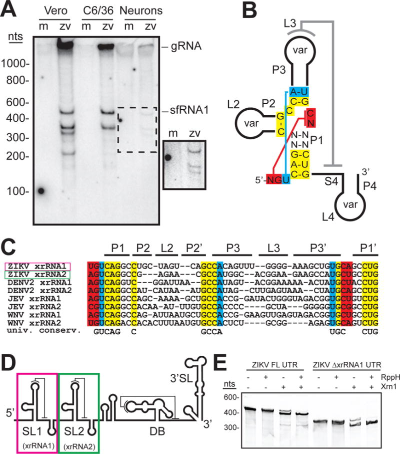
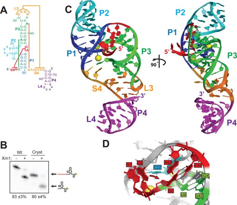
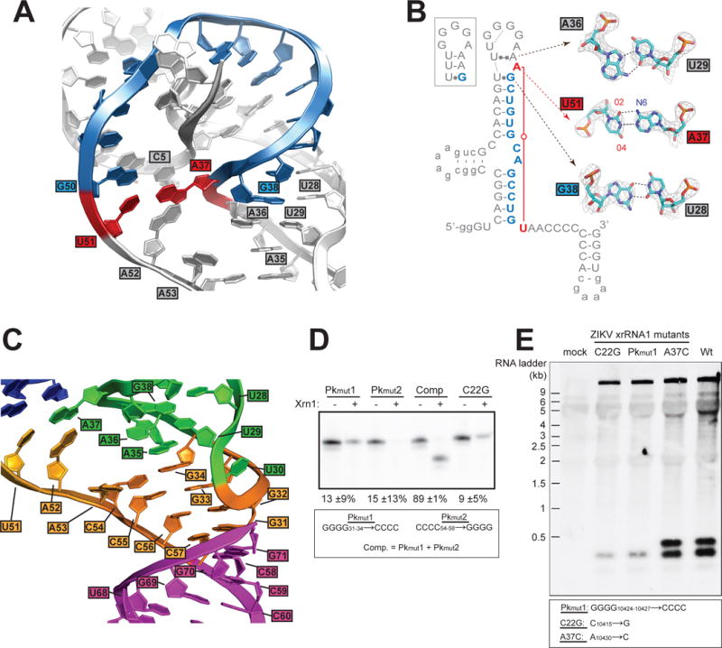
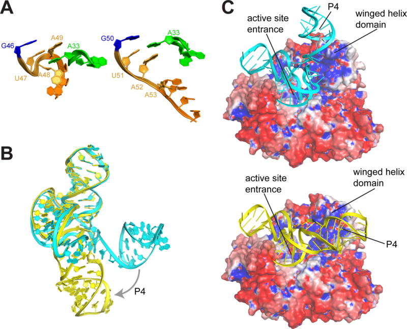
The outbreak of Zika virus (ZIKV) and associated fetal microcephaly mandates efforts to understand the molecular processes of infection. Related flaviviruses produce noncoding subgenomic flaviviral RNAs (sfRNAs) that are linked to pathogenicity in fetal mice. These viruses make sfRNAs by co-opting a cellular exonuclease via structured RNAs called xrRNAs. We found that ZIKV-infected monkey and human epithelial cells, mouse neurons, and mosquito cells produce sfRNAs. The RNA structure that is responsible for ZIKV sfRNA production forms a complex fold that is likely found in many pathogenic flaviviruses. Mutations that disrupt the structure affect exonuclease resistance in vitro and sfRNA formation during infection. The complete ZIKV xrRNA structure clarifies the mechanism of exonuclease resistance and identifies features that may modulate function in diverse flaviviruses.


Figures

Fig. 1
Fig. 1
ZIKV produces sfRNAs by exonuclease resistance. A. Northern blot of purified RNA from mock (m)- and ZIKV (zv)-infected human A549, Vero, mosquito C6/36, and cultured primary mouse neuron cells. Probe was complementary to the putative dumbbell sequence in the ZIKV 3′ UTR. gRNA: genomic RNA. sfRNA1: band corresponding to largest sfRNA. Inset: high-contrast view of the region in the dashed box. Infections and northern blots were confirmed in three independent experiments. B. Secondary structure diagram with sequence conservation in known stem-loop (SL)-type xrRNAs from MbFVs. Universally conserved nucleotides and the position of tertiary interactions are shown. The P2, P3, and P4 stems and associated loops contain variable sequences (var). C. Sequence alignment of several known or putative MbFV xrRNA sequences with regions colored as in panel B. D. Cartoon of the predicted ZIKV 3′ UTR secondary structure, with SL and dumbbell (DB) elements. In WNVKun, the SL1 and SL2 RNAs are xrRNAs (4, 19, 21). E. Ethidium-stained gel of in vitro transcribed ZIKV 3′ UTR or a 3′ UTR lacking the first putative xrRNA1 element (∆xrRNA1) treated with recombinant RppH (to generate a 5′ monophosphate) and recombinant Xrn1. The smaller band indicates Xrn1 resistance. The presence of Xrn1 activity in the absence of RppH is due to spontaneous loss of the 5′ pyrophosphate moiety that occurs at some level even in the absence of the RppH enzyme, as has been previously observed (19).
Fig. 2
Fig. 2
Structure of the ZIKV xrRNA1. A. Secondary structure diagram of the crystallized RNA. Lower case letters represent sequences altered to facilitate RNA expression and crystallization. Colored lines indicate interactions discussed in the text. B. Xrn1 resistance assays of wild-type (Wt) ZIKV xrRNA1 and the crystallized RNA (Cryst) using 3′ end-labeled RNA (yellow circle). The percent of the total RNA that formed an Xrn1-resistant band (listed below the gel) was quantified and reported as the average +/− one standard deviation from the mean of three independent experiments. C. Ribbon representation of the structure of ZIKV xrRNA1, colored to match panel A. Magnesium ions are shown as yellow spheres. D. Detail of interactions at the 5′ end of the RNA. Residue C22 (cyan) contacts the phosphate backbone of neighboring residues, setting up a kink in the RNA critical for folding. Residues U4, A24, and U42 (green) form a base triple interaction orienting the 5′ end. Residue G3 forms a long-range base pairing interaction with residue C44. Residue G2 was mutated from a U to promote transcription; the wild-type sequence is predicted to form a base pair with residue A45 (predicted position change indicated by arrow) (21).
Fig. 3
Fig. 3
Details of the ZIKV xrRNA1 structure. A. Detailed view of the A37-U51 base pair (red) and intervening nucleotides (blue), which circle the 5′ end of the RNA. Other nucleotides discussed in the text are labeled. B. A37-U51 base pair (red) and intervening nucleotides (blue) are highlighted. The box shows the previously predicted secondary structure of the P3-L3 stem-loop. Leontis-Westhof nomenclature is used to indicate non-canonical pairing (30). Inset displays details of all three non-canonical base pairs; electron density is displayed at the 2 sigma contour level. C. The L3–S4 pseudoknot with the P4 stem co-axially stacked. Color matches figure 2A&C. D. Xrn1 resistance assays of pseudoknot mutants and a mutant known to disrupt xrRNA folding (C22G) (19, 21). Quantitation of resistance from three experiments is shown, determined as in figure 2B. E. Northern blot of viral RNA isolated from viral infection with wild-type virus and virus mutated in the xrRNA1 structure. The mutants are labeled to match the analogous mutants in panel D and Figure S4C; corresponding positions in the viral RNA are provided in the box.
Fig. 4
Fig. 4
Model of ZIKV xrRNA-Xrn1 interaction. A. Comparison of the S4 region (orange) and adjacent in the partially folded MVE (left) and fully folded ZIKV (right) xrRNAs. B. Overlay of the MVE (cyan) and ZIKV (yellow) structures, showing the change in the position of the P4/L4 hairpin. C. Models of the MVE (top) and ZIKV (bottom) xrRNAs docked onto the surface of Xrn1 colored by electrostatic potential (blue = positive, red = negative). Structural features are labeled.

Similar articles

Cited by

References

    1. Mackenzie JS, Gubler DJ, Petersen LR. Emerging flaviviruses: the spread and resurgence of Japanese encephalitis, West Nile and dengue viruses. Nature medicine. 2004;10:S98–109. - PubMed
    1. Petersen LR, Jamieson DJ, Powers AM, Honein MA. Zika Virus. The New England journal of medicine. 2016;374:1552–1563. - PubMed
    1. Lindenbach BD, Thiel HJ, Rice CM. In: Fields Virology. 5. Knipe DM, Howley PM, editors. 2007.
    1. Pijlman GP, et al. A highly structured, nuclease-resistant, noncoding RNA produced by flaviviruses is required for pathogenicity. Cell host & microbe. 2008;4:579–591. - PubMed
    1. Manokaran G, et al. Dengue subgenomic RNA binds TRIM25 to inhibit interferon expression for epidemiological fitness. Science. 2015;350:217–221. - PMC - PubMed

Publication types