. 2016 Dec 9:6:38622.
doi: 10.1038/srep38622.

Targeting ASC in NLRP3 inflammasome by caffeic acid phenethyl ester: a novel strategy to treat acute gout

Affiliations

Targeting ASC in NLRP3 inflammasome by caffeic acid phenethyl ester: a novel strategy to treat acute gout

Hye Eun Lee et al. Sci Rep. .

Abstract

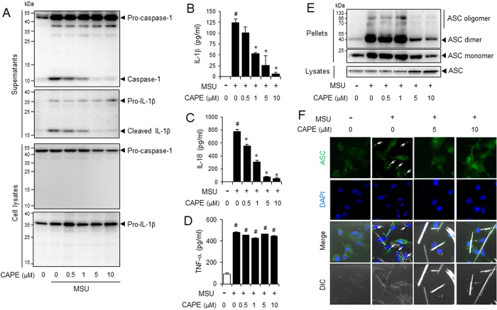
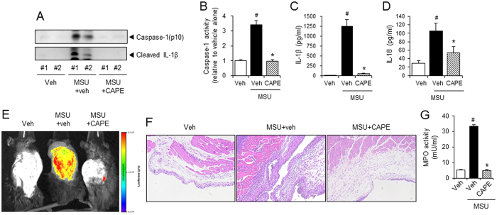
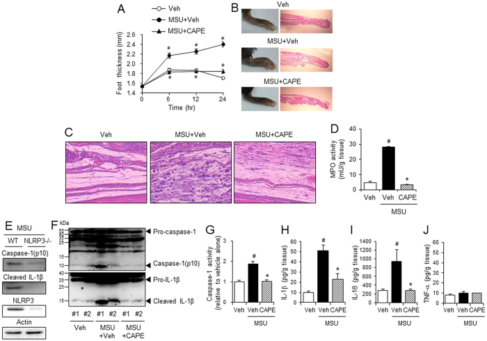
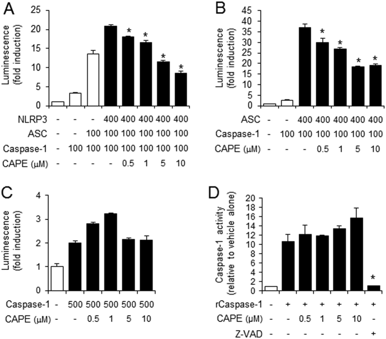
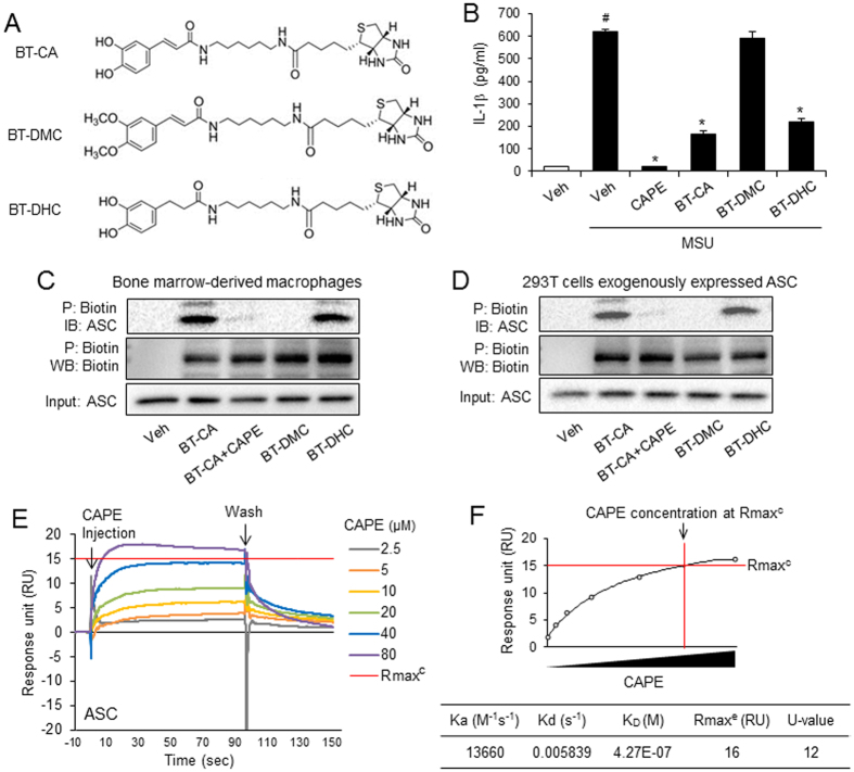
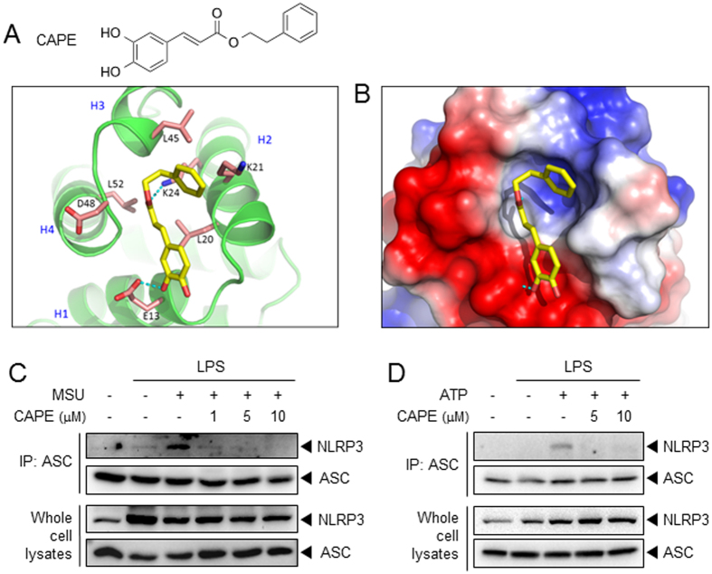
Gouty arthritis is caused by the deposition of uric acid crystals, which induce the activation of NOD-like receptor family, pyrin domain containing 3(NLRP3) inflammasome. The NLRP3 inflammasome, composed of NLRP3, the adaptor protein ASC, and caspase-1, is closely linked to the pathogenesis of various metabolic diseases including gouty arthritis. We investigated whether an orally administrable inhibitor of NLRP3 inflammasome was effective for alleviating the pathological symptoms of gouty arthritis and what was the underlying mechanism. In primary mouse macrophages, caffeic acid phenethyl ester(CAPE) blocked caspase-1 activation and IL-1β production induced by MSU crystals, showing that CAPE suppresses NLRP3 inflammasome activation. In mouse gouty arthritis models, oral administration of CAPE suppressed MSU crystals-induced caspase-1 activation and IL-1β production in the air pouch exudates and the foot tissues, correlating with attenuation of inflammatory symptoms. CAPE directly associated with ASC as shown by SPR analysis and co-precipitation, resulting in blockade of NLRP3-ASC interaction induced by MSU crystals. Our findings provide a novel regulatory mechanism by which small molecules harness the activation of NLRP3 inflammasome by presenting ASC as a new target. Furthermore, the results suggest the preventive or therapeutic strategy for NLRP3-related inflammatory diseases such as gouty arthritis using orally available small molecules.


Figures

Figure 1
Figure 1. CAPE suppresses the MSU crystals-induced activation of the NLRP3 inflammasome in primary macrophages.
Bone marrow-derived macrophages (BMDMs) were primed with LPS (500 ng/ml) for 4 hr. The cells were treated with CAPE for 1 hr and then stimulated with monosodium uric acid (MSU) crystals (500 μg/ml) for (A) 4.5 hr or (BE) 6 hr. In (A), the cell culture supernatants and cell lysates were immunoblotted for pro-caspase-1, caspase-1 (p10), pro-IL-1β, and IL-1β. In (B, C and D) the cell culture supernatants were analyzed for secreted IL-1β, IL-18, and TNF-α using ELISA. The values represent the means ± SEM (n = 3). #Significantly different from vehicle alone, p < 0.05. *Significantly different from MSU alone, p < 0.05. In (E), the cell lysates and crosslinked pellets were resolved using SDS-PAGE and were immunoblotted for ASC. In (F), the cells were fixed, permeabilized and stained for ASC (green), and the nuclei were stained with 4′,6-diamidino-2-phenylindole (DAPI; blue). The arrows indicate ASC speckles. The data are representative of three independent experiments. CAPE, caffeic acid phenethyl ester; MSU, monosodium uric acid crystals. DIC, differential interference contrast.
Figure 2
Figure 2. Oral administration of CAPE attenuates MSU crystals-induced NLRP3 inflammasome activation in a mouse air pouch model.
(A–E) Air pouches were formed on the dorsa of C57BL/6 mice by injecting air twice. The mice were orally administered CAPE (30 mg/kg) or vehicle (Veh, 0.02% DMSO in water). After 1 hr, MSU crystals (3 mg/ml in PBS/mouse) or PBS alone were injected into the air pouches. After 6 hr, the pouch exudates were harvested and the supernatants were analyzed by (A) immunoblotting for caspase-1(p10) and IL-1β, (B) caspase-1 enzyme activity assay, and ELISAs for (C) IL-1β, and (D) IL-18. (E) Bone marrow-derived immortalized macrophages that had been transfected with the iGLuc luciferase reporter plasmid were injected into the air pouches. After 3 hr, the mice were orally administered CAPE (30 mg/kg) or vehicle. After 1 hr, MSU crystals (3 mg/ml in PBS/mouse) or PBS alone were injected into the air pouches. After 6 hr, luminescence derived from iGLuc-luciferase expression was assessed by in vivo imaging analysis using an Xtreme system (Bruker). (F) The air pouch tissue was fixed for histological examination using H&E staining. The purple dots represent infiltrated neutrophils. (G) Myeloperoxidase (MPO) activity, which reflects neutrophil recruitment, was assessed in the air pouch exudates. The values in the bar graphs represent the means ± SEM (n = 3–6 mice). #Significantly different from vehicle alone, p < 0.05. *Significantly different from MSU alone, p < 0.05. Veh, vehicle.
Figure 3
Figure 3. Oral administration of CAPE prevents MSU crystals-induced gout in mouse foot by blocking NLRP3 inflammasome activation.
Mice were orally administered CAPE (30 mg/kg) or vehicle (Veh, 0.02% DMSO in water). After 1 hr, MSU crystals (2 mg/0.1 ml of PBS/mouse) or PBS alone were subcutaneously injected into the pad of the right hind foot of each mouse. After 24 hr, the footpad tissue was collected for analysis. (A) Time course of foot thickness. (B) Representative photographs and H&E staining of the hind feet. (C) Infiltrated neutrophils in the hind foot tissue appear as purple dots in H&E staining (400X). (D) Supernatants from the foot tissue lysates were analyzed for myeloperoxidase (MPO) activity. (E) MSU crystals (2 mg/0.1 ml of PBS/mouse) or PBS alone were subcutaneously injected into the pads of the right hind feet of wild-type (WT) and NLRP3-knockout mice. The foot tissue was analyzed by immunoblotting for caspase-1(p10), IL-1β, NLRP3, and actin. (F–J) The foot tissues from Fig. 3A were subjected to immunoblotting for pro-caspase-1, caspase-1(p10), pro-IL-1β, and IL-1β, a caspase-1 enzyme activity assay, and ELISAs for IL-1β, IL-18, and TNF-α. The values in the line and bar graphs represent the means ± SEM (n = 3 mice). #Significantly different from vehicle alone, p < 0.05. *Significantly different from MSU alone, p < 0.05. Veh, vehicle.
Figure 4
Figure 4. The suppression of inflammasome activation by CAPE is dependent on ASC.
(A–C) 293T cells were transiently transfected with the iGLuc luciferase reporter plasmid (100 ng) and expression plasmids. Luminescence derived from iGLuc activation in each sample was normalized by β-galactosidase activity transfected as an internal control in each sample. (A) *Significantly different from NLRP3 + ASC + caspase-1, 0.5: p = 0.0064, 1–10: p =< 0.0001. (B) *Significantly different from ASC + caspase-1, 0.5: p = 0.0036, 1: p = 0.0001, 5–10: p =< 0.0001. (D) In vitro assay for caspase-1 enzyme activity was performed using a fluorometric caspase-1 assay kit with recombinant human caspase-1 (rCaspase-1; Bio-vision) in the presence or absence of CAPE or Z-VAD-FMK according to the manufacture’s instruction. The fluorescence was recorded at 400 nm after excitation at 505 nm with SpectraMaxM5 (Molecular Devices, Sunnyvale, CA). *Significantly different from rCaspase-1 alone, p = 0.0007. The values represent the means ± SEM (n = 3).
Figure 5
Figure 5. CAPE associated with ASC expressed endogenously and exogenously in the cell.
(A) The structure of biotin-tagged CA (BT-CA), biotin-tagged DMC (BT-DMC), and biotin-tagged DHC (BT-DHC). (B) LPS-primed BMDMs were treated with CAPE, BT-CA, BT-DMC, and BT-DHC (10 μM) for 1 hr and then stimulated with monosodium uric acid (MSU) crystals (500 μg/ml) for 6 hr. The cell culture supernatants were analyzed for secreted IL-1β using ELISA. The values represent the means ± SEM (n = 3). #Significantly different from vehicle alone, p < 0.0001. *Significantly different from MSU alone, p < 0.0001. (C) After BMDM cell lysates were treated with BT-CA, BT-DMC, and BT-DHC (1 μM) at room temperature for 4 hr, cell lysates were precipitated with NeutrAvidin beads and subjected to immunoblotting analysis. The amount of ASC expression in cell lysates were determined as “input”. CAPE (1 μM) was added to cell lysates treated with BT-CA. (D) After 293T cells were transfected with ASC-expression plasmids, the cell lysates were treated with BT-CA, BT-DMC, and BT-DHC (1 μM) at room temperature for 4 hr. The cell lysates were precipitated with NeutrAvidin beads and subjected to immunoblotting analysis. CAPE (1 μM) was added to cell lysates treated with BT-CA. (E) Sensograms of CAPE binding to recombinant ASC protein in the presence of detergent (0.005% Tween-20) were obtained from surface plasmon resonance (SPR) analysis. Different concentrations of CAPE are presented as an overlay plot aligned at the start of injection. (F) The line graph of dose-binding response unit curve and the table showing kinetic parameters of the binding between CAPE and ASC calculated using a simple 1:1 interaction model were from SPR analysis in (E). The maximal expected binding level (Rmaxc) was calculated by Biocore T200 evaluation software and Rmaxe value was obtained from experimental maximum response unit. Veh, vehicle. P, precipitation. IB, immunoblotting. WB, western blotting.
Figure 6
Figure 6. CAPE blocks the interaction between NLRP3 and ASC.
(A) The chemical structure of CAPE and the proposed molecular docking model for CAPE binding to ASC. (B) Electrostatic surface binding model for CAPE and ASC. Red: negative charge, blue: positive charge. (C,D) BMDMs were primed with LPS (C, 500 ng/ml; D, 100 ng/ml) for 4 hr. Then, the cells were treated with CAPE for 1 hr, followed by stimulation with MSU (500 μg/ml) for 5 hr or ATP (5 mM) for 1 hr. Cell lysates were immunoprepitated with anti-ASC antibody followed by immunoblotting as indicated. Representative data from at least two independent experiments are presented.

References

    1. Zhu Y., Pandya B. J. & Choi H. K. Prevalence of gout and hyperuricemia in the US general population: the National Health and Nutrition Examination Survey 2007–2008. Arthritis Rheum 63, 3136–3141, doi: 10.1002/art.30520 (2011). - DOI - PubMed
    1. Sandler D. P., Burr F. R. & Weinberg C. R. Nonsteroidal anti-inflammatory drugs and the risk for chronic renal disease. Ann Intern Med 115, 165–172 (1991). - PubMed
    1. So A., De Smedt T., Revaz S. & Tschopp J. A pilot study of IL-1 inhibition by anakinra in acute gout. Arthritis Res Ther 9, R28, doi: 10.1186/ar2143 (2007). - DOI - PMC - PubMed
    1. Dumusc A. & So A. Interleukin-1 as a therapeutic target in gout. Curr Opin Rheumatol 27, 156–163, doi: 10.1097/bor.0000000000000143 (2015). - DOI - PubMed
    1. McCarty D. J. & Hollander J. L. Identification of urate crystals in gouty synovial fluid. Ann Intern Med 54, 452–460 (1961). - PubMed

Publication types

MeSH terms