. 2017 Jan;93(1):343-354.
doi: 10.1111/php.12684.

An Ethenoadenine FAD Analog Accelerates UV Dimer Repair by DNA Photolyase

Affiliations

An Ethenoadenine FAD Analog Accelerates UV Dimer Repair by DNA Photolyase

Madhavan Narayanan et al. Photochem Photobiol. 2017 Jan.

Erratum in

Abstract

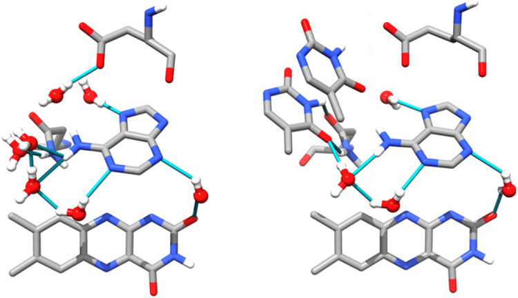
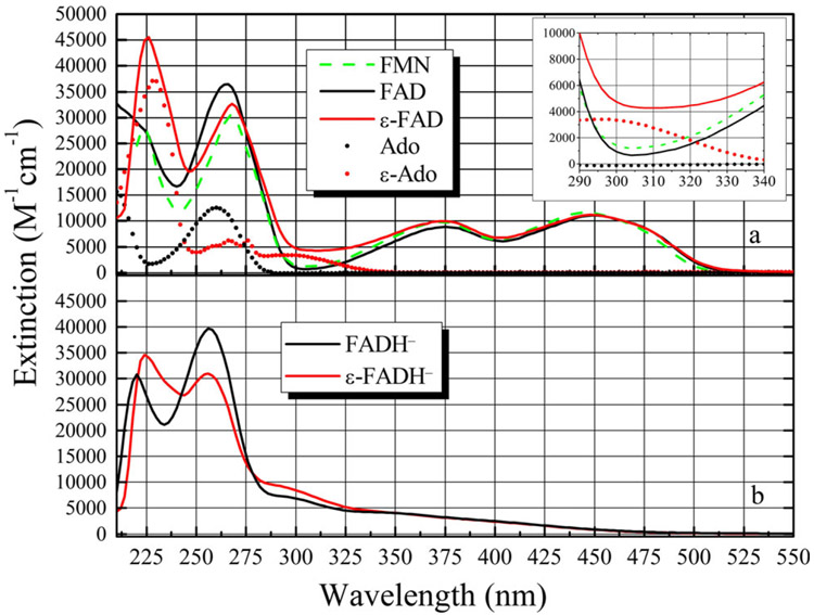
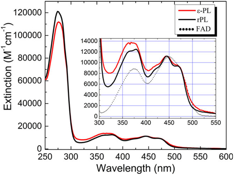
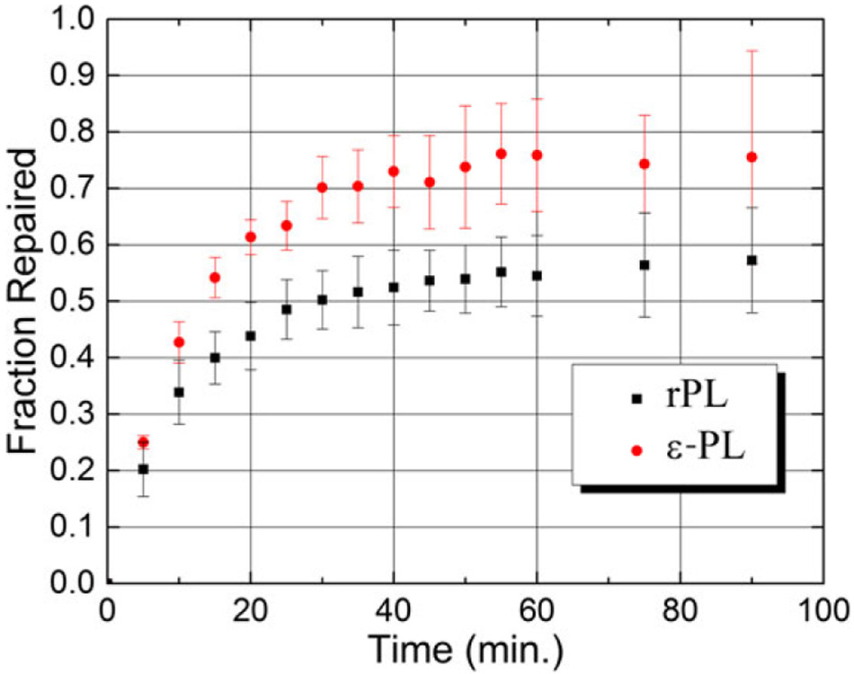
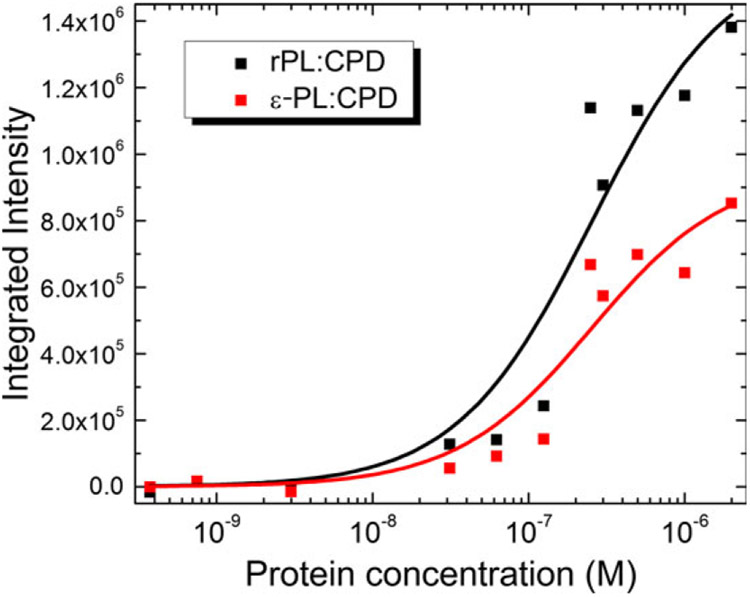
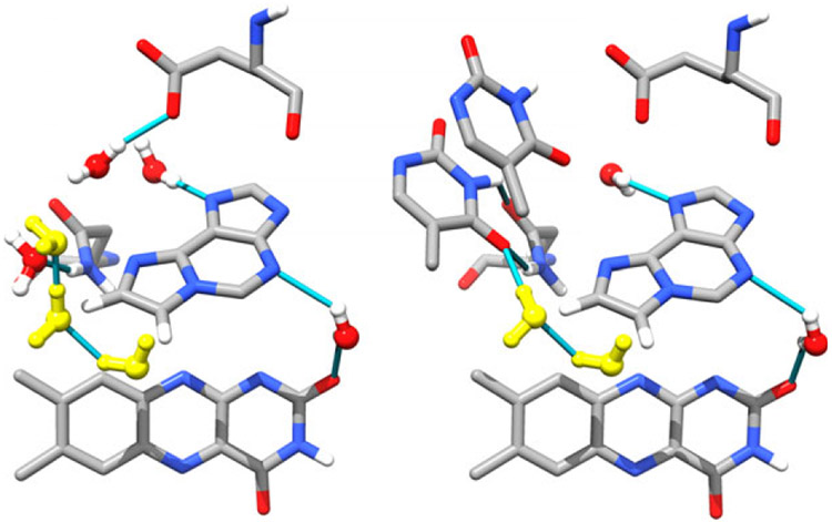
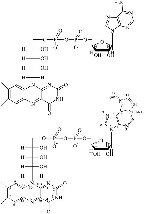
Reduced anionic flavin adenine dinucleotide (FADH- ) is the critical cofactor in DNA photolyase (PL) for the repair of cyclobutane pyrimidine dimers (CPD) in UV-damaged DNA. The initial step involves photoinduced electron transfer from *FADH- to the CPD. The adenine (Ade) moiety is nearly stacked with the flavin ring, an unusual conformation compared to other FAD-dependent proteins. The role of this proximity has not been unequivocally elucidated. Some studies suggest that Ade is a radical intermediate, but others conclude that Ade modulates the electron transfer rate constant (kET ) through superexchange. No study has succeeded in removing or modifying this Ade to test these hypotheses. Here, FAD analogs containing either an ethano- or etheno-bridged Ade between the AN1 and AN6 atoms (e-FAD and ε-FAD, respectively) were used to reconstitute apo-PL, giving e-PL and ε-PL respectively. The reconstitution yield of e-PL was very poor, suggesting that the hydrophobicity of the ethano group prevented its uptake, while ε-PL showed 50% reconstitution yield. The substrate binding constants for ε-PL and rPL were identical. ε-PL showed a 15% higher steady-state repair yield compared to FAD-reconstituted photolyase (rPL). The acceleration of repair in ε-PL is discussed in terms of an ε-Ade radical intermediate vs superexchange mechanism.


Figures

Figure 1.
Figure 1.
The active site of DNA photolyase without (left, ITEZ.PDB (18), chain C) and with substrate (right, ITEZ.PDB (18), chain A). The adenine and CPD are shown as sticks. The isoalloxazine ring is shown in wire form. The conserved water molecules are shown in red and white balls and sticks with hydrogen bonds shown as blue lines. Glu283 and Asn349, which make contacts with the CPD and/or Ade. In the PL:CPD complex, Ade is in vdW contact with the CPD. The ribityl-phosphate linker has been removed for clarity.
Figure 2.
Figure 2.
(a) Absorption spectra of FAD (–––), FMN (–––) and ε-FAD (–––). Also shown are Ade (●) and ε-Ade () for comparison. The inset shows the region from 290 to 340 nm where the flavin molecules have a minimum in their absorbance, while the ε-Ade group has roughly 3 times the extinction at ca. 305 nm. (b) Photoreduction of ε-FAD→ε-FADH does not reduce the ε-Ade molecule, as shown by the unchanged spectral band around 305 nm. FADH is also shown for comparison.
Figure 3.
Figure 3.
Absorption spectrum of FAD (rPL) and ε-FAD (ε-PL) reconstituted photolyase. The spectra were taken in buffer Apo-A (which includes 1.7 m ammonium sulfate) and normalized to 11 200 m−1 cm−1 at 444 nm. The flavin vibronic features are clearly seen in the S0→S2 band around 444 nm. Inset: The S0→S1,2 bands of ε-PL show a small degree of broadening compared to the rPL spectrum. This suggests that the modified cofactor is not as rigidly bound as the FAD.
Figure 4.
Figure 4.
The fraction of repaired T5 substrate with irradiation time for rPL and ε-PL. The error bars represent the standard error from the mean derived from three replicates. The repair yield of ε-PL is about 15% higher than rPL.
Figure 5.
Figure 5.
Plots of integrated fluorescence emission vs. protein concentration against a fixed concentration of substrate (250 nm). Protein emission sans substrate was subtracted at each concentration point. The emission data were then integrated from 359 to 387 nm to exclude emission from the ε-Ade chromophore. KA for rPL was (3.9 ± 1.4) × 106 m−1 and that for ε-PL was (4.0 ± 1.5) × 106 m−1, as obtained from a fit to a hyperbolic function (solid lines).
Figure 6.
Figure 6.
Left. Cartoon of ε-FAD structure inside of apo-PL constructed using Chimera (UCSF (75)). Water molecules that are no longer hydrogen bonded to adenine are shown in yellow and are probably lost due to steric clashes from the ε-Ade group. Right: Chimera cartoon of how the ε-Ade group eliminates important contacts with the CPD, while increasing orbital overlap with the flavin ring.
Scheme 1.
Scheme 1.
Structures of FAD and etheno-FAD (ε-FAD), showing the differences in the adenine and ε-Ade structures. Numbering of the adenines is based on the purine system (77).

References

    1. Taylor J-S (1994) Unraveling the molecular pathway from sunlight to skin cancer. Acc. Chem. Res 27, 76–82.
    1. Kao Y-T, Saxena C, He T-F, Guo L, Wang L, Sancar A and Zhong D (2008) Ultrafast dynamics of flavins in five redox states. J. Am. Chem. Soc 130, 13132–13139. - PMC - PubMed
    1. LeClerc JE, Borden A and Lawrence CW (1991) The thymine-thymine pyrimidine-pyrimidone (6-4) ultraviolet light photoproduct is highly mutagenic and specifically induces 3’ thymine-to-cytosine transitions in Escherichia coli. Proc. Natl Acad. Sci. USA 88, 9685–9689. - PMC - PubMed
    1. Sancar A, Franklin KA and Sancar GB (1984) Escherichia coli DNA photolyase stimulates uvrABC excision nuclease in vitro. Proc. Natl Acad. Sci. USA 81, 7397–7401. - PMC - PubMed
    1. Minato S and Werbin H (1971) Spectral properties of the chromophoric material associated with the deoxyribonucleic acid photoreactivating enzyme isolated from baker’s yeast. Biochemistry 10, 4503–4508. - PubMed

Publication types

LinkOut - more resources