. 2017 Jan 3;8(1):1354-1368.
doi: 10.18632/oncotarget.13817.

The involvement of Bcl-2 family proteins in AKT-regulated cell survival in cisplatin resistant epithelial ovarian cancer

Affiliations

The involvement of Bcl-2 family proteins in AKT-regulated cell survival in cisplatin resistant epithelial ovarian cancer

Yan Dai et al. Oncotarget. .

Erratum in

Abstract

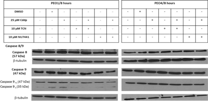
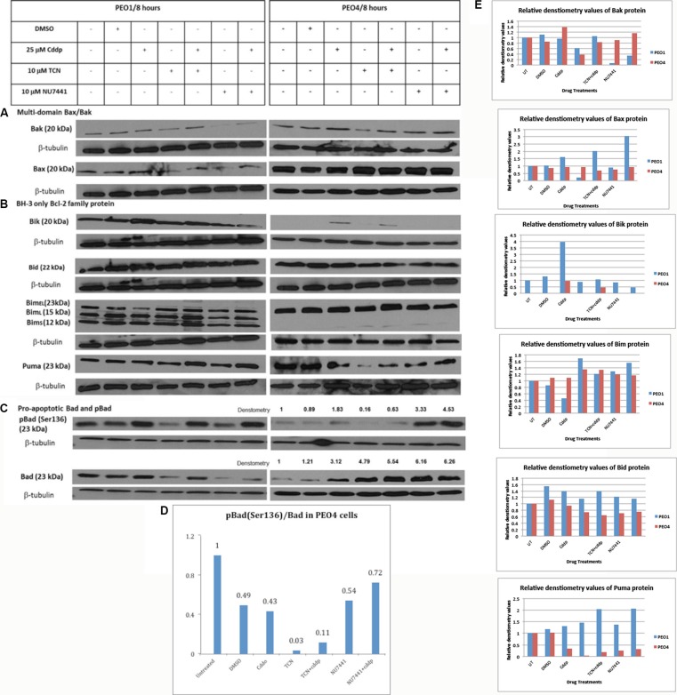
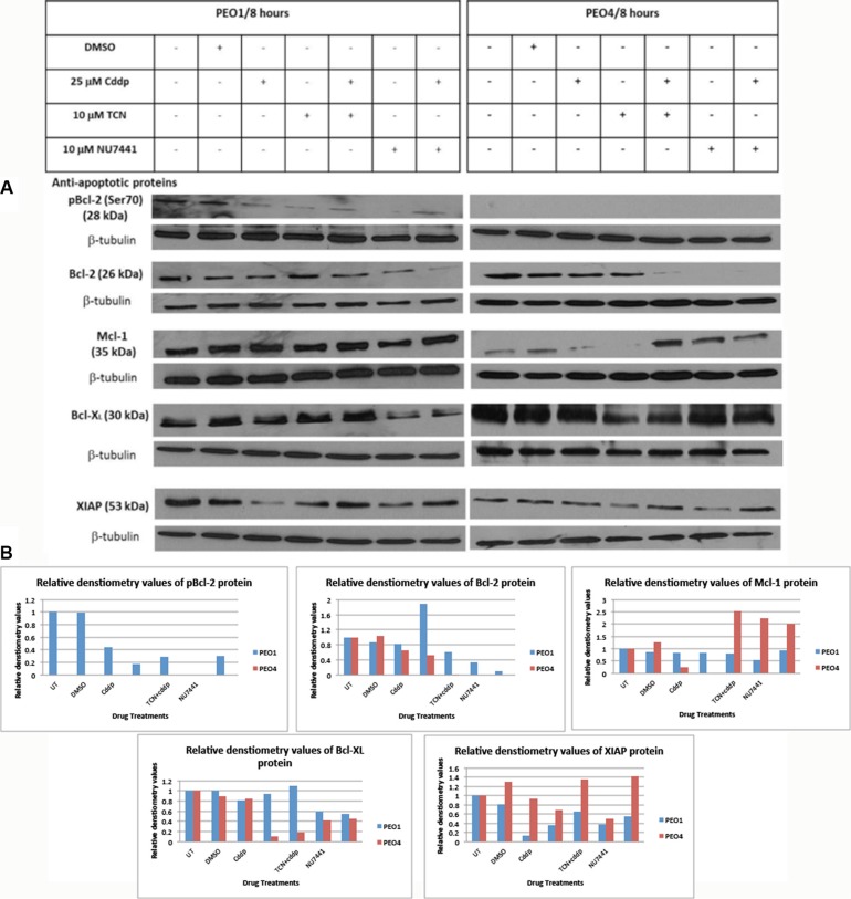
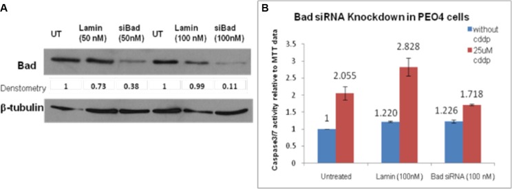
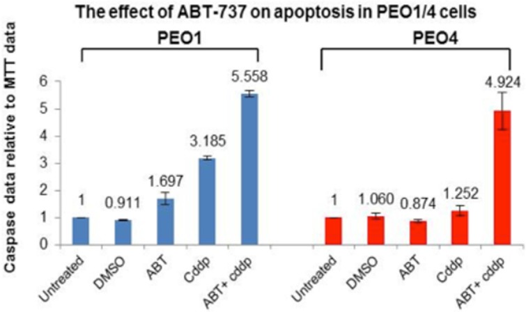
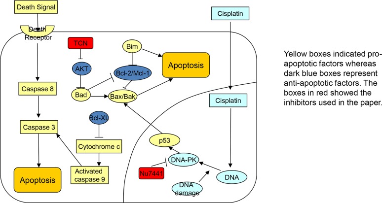
Many studies involving patients with cisplatin-resistant ovarian cancer have shown that AKT activation leads to inhibition of apoptosis. The aim of this study was to examine the potential involvement of the Bcl-2 family proteins in AKT-regulated cell survival in response to cisplatin treatment. Cisplatin-sensitive (PEO1) and cisplatin-resistant (PEO4) cells were taken from ascites of patients with ovarian cancer before cisplatin treatment and after development of chemoresistance. It was found that cisplatin treatment activated the AKT signaling pathway and promoted cell proliferation in cisplatin-resistant EOC cells. When AKT was transfected into nucleus of cisplatin-resistant ovarian cancer cells, DNA-PK was phosphorylated at S473. The activated AKT (pAKT-S473) in these cells inhibited the death signal induced by cisplatin thereby inhibiting cisplatin-mediated apoptosis. Results from this study showed that the combination of cisplatin, DNA-PK inhibitor NU7441, and AKT inhibitor TCN can overcome drug resistance, increase apoptosis, and re-sensitize PEO4 cells to cisplatin treatment. A decrease in apoptotic activity was seen in PEO4 cells when Bad was downregulated by siRNA, which indicated that Bad promotes apoptosis in PEO4 cells. Use of the Bcl-2 inhibitor ABT-737 showed that ABT-737 binds to Bcl-2 but not Mcl-1 and releases Bax/Bak which leads to cell apoptosis. The combination of ABT-737 and cisplatin leads to a significant increase in the death of PEO1 and PEO4 cells. All together, these results indicate that Bcl-2 family proteins are regulators of drug resistance. The combination of cisplatin and Bcl-2 family protein inhibitor could be a strategy for the treatment of cisplatin-resistant ovarian cancer.

Keywords: AKT; Bcl-2; cisplatin; drug resistance; epithelial ovarian cancer.


Conflict of interest statement

CONFLICTS OF INTERESTS

Authors declare no conflicts of interests.

Figures

Figure 1
Figure 1. Expressions of caspase 8 and caspase 9 proteins in PEO1 and PEO4 cells in response to treatment with cisplatin, TCN and NU7441
Cisplatin-sensitive PEO1 cells and cisplatin-resistant PEO4 cells were treated as indicated. Cell lysates were prepared and were resolved by 12% SDS-PAGE agarose gel as described in Materials and Methods. The blots were probed with anti-caspase 8/9 antibodies and all blots were re-probed for b-tubulin expression as a loading control.
Figure 2
Figure 2. Expression of pro-apoptotic Bcl-2 family proteins in PEO1 and PEO4 cells in response to treatment with cisplatin, TCN and NU7441
Cisplatin-sensitive PEO1 and cisplatin—resistant PEO4 cells were treated as indicated. Cell lysates were prepared and were separated by 12% SDS-PAGE gel as described in Materials and Methods. Western Blotting was carried out to examine expression of (A) multi-domain pro-apoptotic Bax and Bak (B) pro-apoptotic BH-3 only Bcl-2 family proteins (C) total and phosphorylated pro-apoptotic Bad. (D and E) Densitometry values for protein expression in PEO4 cells analyzed by Image J software and normalized to their corresponding b-tubulin densities.
Figure 3
Figure 3. Expression of anti-apoptotic Bcl-2 family proteins in PEO1 and PEO4 cells in response to treatment with cisplatin, TCN and NU7441
Cisplatin-sensitive PEO1 and cisplatin-resistant PEO4 cells were treated as indicated. Cell lysates were prepared and were separated by 12% SDS-PAGE gel as described in Materials and Methods. (A) Blots were probed with anti-apoptotic pBcl-2 (Ser70), total Bcl-2, Mcl-1, Bcl-XL, and XIAP and all membranes were incubated with β-tubulin, which was used as a loading control. (B) Densitometry values for protein expression in PEO4 cells analyzed by Image J software and normalized to their corresponding β-tubulin densities.
Figure 4
Figure 4
(A) siRNA knockdown of Bad in PEO4 cells. PEO4 cells were treated with Bad siRNA or Lamin control siRNA, and expression of total Bad were assessed by Western Blotting as described in Materials and Methods. 90% knockdown of Bad was achieved when PEO4 cells were transfected with Bad siRNA (100 nM). The apoptotic activity induced by cisplatin was significantly reduced by Bad siRNA when compared to that in untreated cells. (B) Bad siRNA knockdown repressed the apoptotic activity of cisplatin in chemo-resistant PEO4 cells. The caspase3/7 assay and MTT assay were performed to observe the influence of Bad knockdown on apoptosis in response to 25 μM cisplatin treatment. siGenome Lamin A/C control was used as an experimental control and the Lamin transfection did not affect the apoptotic response of PEO4 cells to cisplatin.
Figure 5
Figure 5. The effect of ABT-737 inhibitor on cisplatin-induced apoptosis in PEO1 and PEO4 cells
A small molecule BH-3 mimetic inhibitor ABT-737 (1 μM), which specifically inhibits Bcl-2 and Bcl-XL, was used to treat PEO1 and PEO4 cells individually or in combination with 25 μM cddp. After 8-hr incubation, ABT-737 inhibitor increased the chemo-sensitivity of PEO1 and PEO4 cells to cddp to a high level compared to their corresponding basal levels (25 μM cddp).
Figure 6
Figure 6. Schematic picture
The signaling pathways related to combination treatment using cisplatin, DNA-PK inhibitor NU7441, and AKT inhibitor TCN.

References

    1. Cho KR, Shih IeM. Ovarian cancer. Annu Rev Pathol. 2009;4:287–313. - PMC - PubMed
    1. Agarwal R, Kaye SB. Ovarian cancer: strategies for overcoming resistance to chemotherapy. Nat Rev Cancer. 2003;3:502–16. - PubMed
    1. Lokadasan R, James FV, Narayanan G, Prabhakaran PK. Targeted agents in epithelial ovarian cancer: review on emerging therapies and future developments. E cancer medical science. 2016;10:626. - PMC - PubMed
    1. Bozulic L, Surucu B, Hynx D, Hemmings BA. PKB alpha/Akt1 acts downstream of DNA-PK in the DNA double-strand break response and promotes survival. Mol Cell. 2008;30:203–13. - PubMed
    1. Fraser M, Bai T, Tsang BK. Akt promotes cisplatin resistance in human ovarian cancer cells through inhibition of p53 phosphorylation and nuclear function. Int J Cancer. 2008;122:534–46. - PubMed

MeSH terms