. 2016 Dec 9;11(12):e0167116.
doi: 10.1371/journal.pone.0167116. eCollection 2016.

A Cost-Effective Method to Assemble Biomimetic 3D Cell Culture Platforms

Affiliations

A Cost-Effective Method to Assemble Biomimetic 3D Cell Culture Platforms

Sabreen Khalil et al. PLoS One. .

Abstract

Methods: We utilized the hAM to provide the biological and the three dimensional (3D) topographic components of the prototype. The 3D nano-roughness of the hAM was characterized using surface electron microscopy and surface image analysis (ImageJ and SurfaceJ). We developed additional macro-scale and micro-scale versions of the platform which provided additional shear stress factors to simulate the fluid dynamics of the in vivo extracellular fluids.

Results: Three models of varying complexities of the prototype were assembled. A well-defined 3D surface modulation of the hAM in comparable to commercial 3D biomaterial culture substrates was achieved without complex fabrication and with significantly lower cost. Performance of the prototype was demonstrated through culture of primary human umbilical cord mononuclear blood cells (MNCs), human bone marrow mesenchymal stem cell line (hBMSC), and human breast cancer tissue.

Conclusion: This study presents methods of assembling an integrated, flexible and low cost biomimetic cell culture platform for diverse cell culture applications.


Conflict of interest statement

We have declared in our first submission a filed patent related to this work. The patent was filed in June, 2015 as PCT/EG2015/000030 and has not been published yet. We confirm that this does not alter our adherence to PLOS ONE policies on sharing data and materials.

Figures
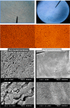
Fig 1
Fig 1. Human Amniotic Membrane Surface Treatment.
This figure shows the fetal side of the hAM in its intact (left column) and decellularized (right column) states. Complete decellularization was achieved in repeat experiments using the brief alkaline method as demonstrated by various imaging modalities and magnifications (Top lane shows bright field microscopy images where hAM stained with methylene blue, second lane shows inverted microscopy images followed by SEM images at x500 and x2000).
Fig 2
Fig 2. Prototypes of the Biomimetic Platform.
Various examples of the prototypes are shown. P1 and P2 are platforms in which regular Petri dishes or well plates lined with hAM were used. P3 shows successive steps of developing a PDMS-hAM platform with an additional fluid dynamic factor (a-d). P4 is a microchip chamber connected with microfluidic channels (inlet and outlet channels), the hAM layer was applied top-down to allow the free flow of media across the chamber.
Fig 3
Fig 3. 3D Nano-topography of the Decellularized hAM Based Platform.
The left panel of this Fig shows a SEM image of the hAM at x5000 magnification. It demonstrates the nano-scale topography of the membrane after decellularization. Below the original SEM image, a 3D- surface plot is obtained by ImageJ which illustrates the peaks and valleys distributed homogenously throughout the surface. On the right panel, 3 examples of synthetic scaffolds are presented along with their corresponding 3D surface plotting using the same ImageJ surface plotting parameters. (A) demonstrates a commercial 3D culture plate coating substrate composed of Polytetrafluoroethylene (PTFE) polymer modified with an amphiphilic polymer and collagen. (B) shows a SEM image of a 3D commercial scaffold (at x15000 magnification) composed of inert hydroxypropyl cellulose to form a sponge like structure. (C) demonstrates a synthetic scaffold fabricated from polypyrrole (PPy) and poly(styrene sulfonate) (PSS) interwoven within a PCL matrix[43]
Fig 4
Fig 4. Surface Characterization of the Decellularized hAM Based Platform.
A line diagram illustration of the 3D surface roughness parameter (R) of each scaffold characterized using SurfaceJ. It shows the span between the lowest valley (1), and the highest peak (2) in each scaffold. Detailed SurfaceJ characterization is shown in supplementary information (S1 File).
Fig 5
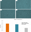
Fig 5. Human Umbilical Cord Blood Mononuclear Cell (MNCs) Culture.
This figure shows progressive proliferation of (MNCs) obtained from the umbilical cord blood and cultured on a 6-well plate lined with hAM (Prototype 1). D0 stands first day immediately after culture. D3, 4, 5 represent cell proliferation on days 3, 4 and 5 respectively. The lower panel shows a bar chart for cell proliferation of the MNCs, in another run of experiments, after 7 days of culture in the hAM coated and non-coated plates. The grey bar represents the original number of cells per ml before culture. (Cell images were obtained by an inverted microscope (x40). (Cropping and coloring of image was slightly done for aesthetic purposes without affecting presented data)
Fig 6
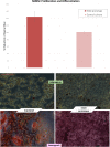
Fig 6. Proliferation and Differentiation of Human Bone Marrow Stem Cells (hBMSC) in the Biomimetic Platform.
The top panel shows a graph depicting proliferation of the hBMSCs as expressed by the relative % reduction of the Alamar Blue reagent using the absorbance values in the study group compared to the control group. The lower panel presents inverted microscope images (x10) of adipogenic (top) and osteogenic differentiation (bottom) of hBMSC grown on prototype 1(hAM coated plates) and control culture plate.

References

    1. Ravi M, Paramesh V, Kaviya SR, Anuradha E, Solomon FD. 3D cell culture systems: advantages and applications. Journal of cellular physiology. 2015;230(1):16–26. Epub 2014/06/10. 10.1002/jcp.24683 - DOI - PubMed
    1. Walasek MA, van Os R, de Haan G. Hematopoietic stem cell expansion: challenges and opportunities. Annals of the New York Academy of Sciences. 2012;1266:138–50. Epub 2012/08/21. 10.1111/j.1749-6632.2012.06549.x - DOI - PubMed
    1. Eggenhofer E, Benseler V, Kroemer A, Popp FC, Geissler EK, Schlitt HJ, et al. Mesenchymal stem cells are short-lived and do not migrate beyond the lungs after intravenous infusion. Frontiers in immunology. 2012;3:297 Epub 2012/10/12. PubMed Central PMCID: PMC3458305. 10.3389/fimmu.2012.00297 - DOI - PMC - PubMed
    1. Scadden David T. Nice Neighborhood: Emerging Concepts of the Stem Cell Niche. Cell. 2014;157(1):41–50. 10.1016/j.cell.2014.02.013 - DOI - PMC - PubMed
    1. Plaks V, Kong N, Werb Z. The Cancer Stem Cell Niche: How Essential Is the Niche in Regulating Stemness of Tumor Cells? Cell Stem Cell. 2015;16(3):225–38. 10.1016/j.stem.2015.02.015 - DOI - PMC - PubMed

MeSH terms

LinkOut - more resources