. 2016 Dec 9;11(12):e0167789.
doi: 10.1371/journal.pone.0167789. eCollection 2016.

Immunolocalization of a Histidine-Rich Epidermal Differentiation Protein in the Chicken Supports the Hypothesis of an Evolutionary Developmental Link between the Embryonic Subperiderm and Feather Barbs and Barbules

Affiliations

Immunolocalization of a Histidine-Rich Epidermal Differentiation Protein in the Chicken Supports the Hypothesis of an Evolutionary Developmental Link between the Embryonic Subperiderm and Feather Barbs and Barbules

Lorenzo Alibardi et al. PLoS One. .

Abstract

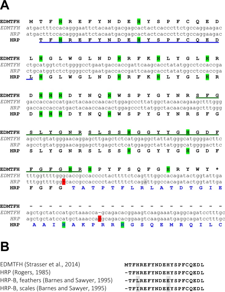
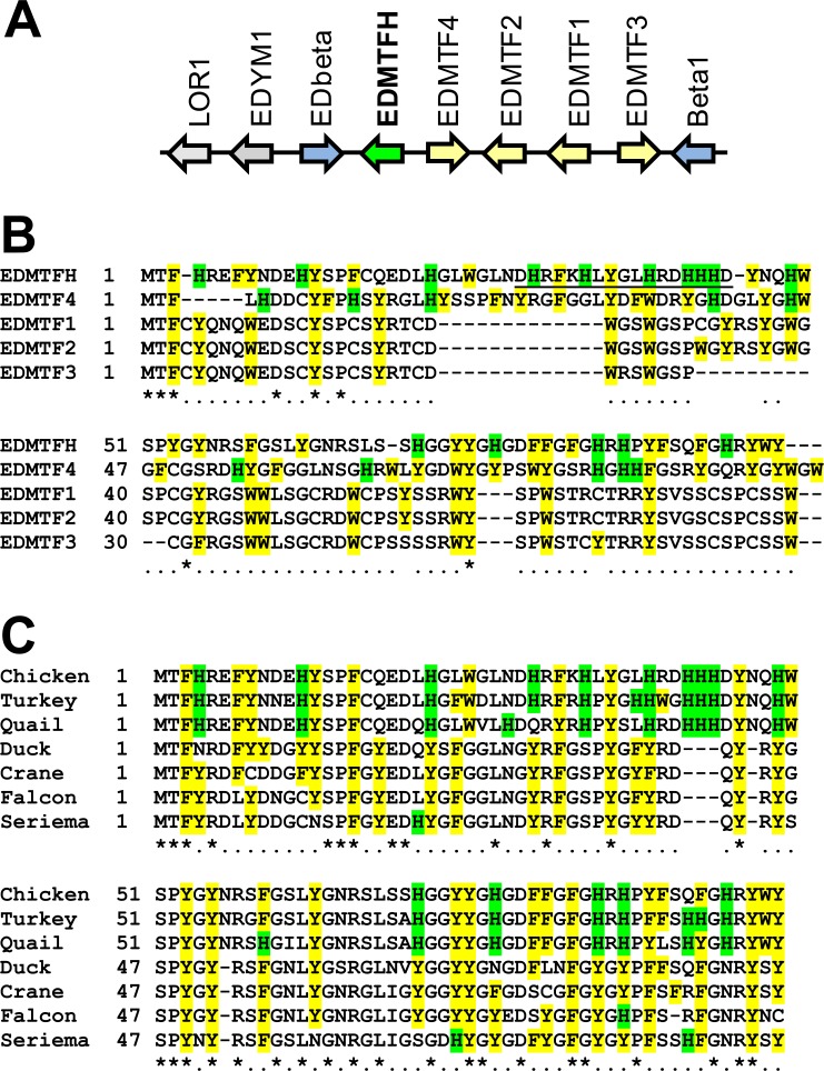
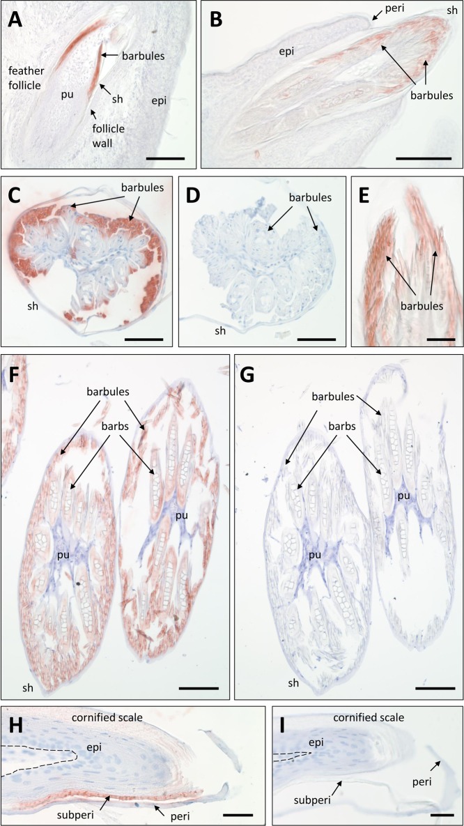
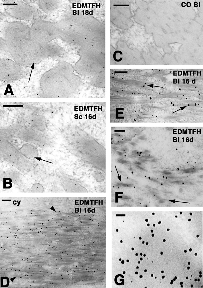
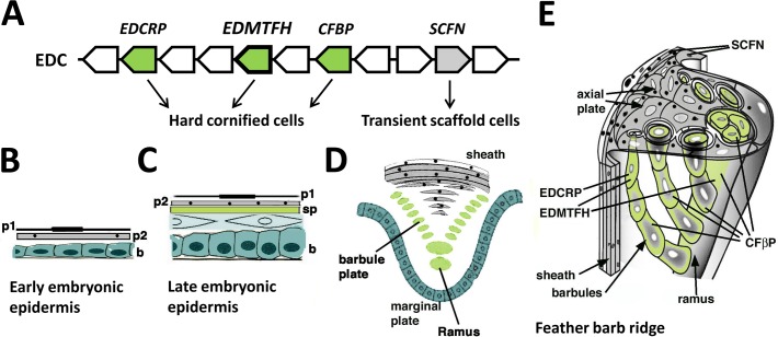
The morphogenesis of feathers is a complex process that depends on a tight spatiotemporal regulation of gene expression and assembly of the protein components of mature feathers. Recent comparative genomics and gene transcription studies have indicated that genes within the epidermal differentiation complex (EDC) encode numerous structural proteins of cornifying skin cells in amniotes including birds. Here, we determined the localization of one of these proteins, termed EDMTFH (Epidermal Differentiation Protein starting with a MTF motif and rich in Histidine), which belongs to a group of EDC-encoded proteins rich in aromatic amino acid residues. We raised an antibody against an EDMTFH-specific epitope and performed immunohistochemical investigations by light microscopy and immunogold labeling by electron microscopy of chicken embryos at days 14-18 of development. EDMTFH was specifically present in the subperiderm, a transient layer of the embryonic epidermis, and in barbs and barbules of feathers. In the latter, it partially localized to bundles of so-called feather beta-keratins (corneous beta-proteins, CBPs). Cells of the embryonic periderm, the epidermis proper, and the feather sheath were immunonegative for EDMTFH. The results of this study indicate that EDMTFH may contribute to the unique mechanical properties of feathers and define EDMTFH as a common marker of the subperiderm and the feather barbules. This expression pattern of EDMTFH resembles that of epidermal differentiation cysteine-rich protein (EDCRP) and feather CBPs and is in accordance with the hypothesis that a major part of the cyclically regenerating feather follicle is topologically, developmentally and evolutionarily related to the embryonic subperiderm.


Conflict of interest statement

The authors have declared that no competing interests exist.

Figures

Fig 1
Fig 1. Nucleotide and amino acid sequence alignments of EDMTFH versus histidine-rich protein (HRP).
(A) The nucleotide sequences of the coding region of chicken EDMTFH [13] and of the chicken HRP cDNA reported previously [29] were aligned. Translations into amino acid sequences are shown above and below the sequences, respectively. Note that insertions and deletions (red shading) in the cDNA sequence relative to the chicken EDMTFH gene in the current genome assembly cause reading frameshifts leading to the prediction of a different carboxy-terminus of HRP (blue fonts) relative to EDMTFH. Sequences corresponding to peptides that were previously identified in feather extracts are marked underlined feather proteins peptides (underlined) corresponding to EDMTFH were identified by (blue underline [29], green underlines [13]). Histidine (H) residues are highlighted by green shading. The stop codon of EDMTFH is marked with an asterisk. (B) Alignment of amino-terminal amino acid sequences of EDMTFH [13] and HRP, as determined by direct sequencing of proteins isolated from feathers [29, 38]. Predicted HRP-B residues that deviate from the EDMTFH sequence at positions of histidines (H) are shaded grey.
Fig 2
Fig 2. Comparison of chicken EDMTFH versus other EDMTF proteins of the chicken and homologs in other species.
(A) EDMTFH gene locus in the chicken. Arrows indicate the origientation of gene transcription. The genes EDbeta and Beta1 (preliminary names [13]) encode corneous beta-proteins (CBPs), also known as beta-keratins. (B) Amino acid sequence alignment of EDMTFH versus other chicken EDMTF proteins. The peptide used as an epitope for raising the anti-EDMTFH antibody is underlined. (C) Amino acid sequence alignment of chicken EDMTFH versus the most similar proteins of other birds. Formats in (B) and (C) indicate the following: Histidine (H) residues are highlighted by green shading, other aromatic residues are highlighted by yellow shading. Numbers indicate amino acid sequence positions. Identity of residues in all sequences is indicated by an asterisk and conservation in at least 50% of the sequences is indicated by "." below the alignments. Hyphens were introduced to maximize the alignment of the sequences. Sequences of EDMTFH orthologs of turkey (Meleagris gallopavo) and quail (Coturnix japonica) were predicted from genomic DNA. Accession numbers of other EDMTFH sequences: AHA62422.1 (chicken, Gallus gallus), XP_012964640.1 (duck, Anas platyrhynchos), XP_010299618.1 (crane, Balearica regulorum gibbericeps), XP_013153676.1 (falcon, Falco peregrinus), XP_009701209.1 (seriema, Cariama cristata).
Fig 3
Fig 3. Light microscopic immunohistochemistry of EDMTFH.
(A-C, E, F) Feather follicles and feathers on the wings of chick embryos on day E18 of development were immunostained with anti-EDMTFH (red). In negative control experiments the primary antibody was replaced with an antiserum raised against an unrelated peptide (D) or preabsorbed with the antigen (G). Scutate scales on the legs of chick embryos (day E19) were immunolabeled for EDMTFH (H) or subjected to the negative control experiment (I). The dermo-epidermal junction is indicated by a dashed line (H, I). epi, epidermis; peri, periderm; pu, pulp; sh, feather sheath; subperi, subperiderm. Bars: 100 μm (A, B, F, G), 50 μm (C-E), 25 μm (H, I).
Fig 4
Fig 4. Ultrastructural localization of EDMTFH by immunogold labeling.
Downfeathers and scales of chicken embroys at days 16 (16d) and 18 (18d) of development were labeled for EDMTFH either without (A-C, G) and with (D-F) silver enhancement. (A) Diffuse labeling over corneous bundles (arrow) of barbule cells (bl). (B) Diffuse labeling in the corneous bundles (arrow) of a subperiderm cell in a scale. (C) Immuno-negative control section of a barbule. (D) Labeling cytoplasmic corneous bundles (arrowheads) but not the cytoplasm (cy) in a barbule cell (bl). (E) Close-up to show the association of the labeling with corneous bundles (arrows). (F) Early differentiating barbule cell with short corneous bundles (arrows). (G) Double-labeling for EDMTFH (5 nm gold particles) and feather beta-keratin (20 nm gold particles) in a barbule cell. Note that the large particles appear to be more abundant than the small particles. A lower magnification image of the double-labeling is shown in S3 Fig. Bars: 100 nm (A, B); 200 nm (C-F); 50 nm (G).
Fig 5
Fig 5. Schematic representation of the contribution of EDMTFH to feather cornification.
(A) EDMTFH and other genes, that encode proteins of the feather follicle, are located in the same gene cluster, the avian epidermal differentiation complex (EDC). EDMTFH, EDCRP and corneous feather beta proteins (CFBPs) are components of hard cornified cells whereas scaffoldin (SCFN) is a component of cells that form a transient scaffold of growing feathers. (B-D) The embryonic epidermis of birds increases the number of layers during development. During late development, 2 layers of periderm (p1, p2) and a subperiderm (sp) are present above the definitive epidermis (C). The feather barb ridge has a topologically similar organization as late embryonic epidermis on scutate scales whereby the equivalents of the periderm form the feather sheath and the axial plate, and barbules cells and rami are equivalent to the subperiderm (D), as suggested by common expression of protein markers. A 3-dimensional depiction of a growing barb ridge of a down feather indicates the different roles of cornifying cells (green) and transient scaffolding cells (grey) in the morphogenesis of feathers (E). Periderm granules containing scaffoldin are indicated as black dots.

References

    1. Widelitz RB, Jiang TX, Yu M, Shen T, Shen JY, Wu P, et al. Molecular biology of feather morphogenesis: a testable model for evo-devo research. J Exp Zool B Mol Dev Evol. 2003;298:109–122. 10.1002/jez.b.29 - DOI - PMC - PubMed
    1. Alibardi L. Adaptation to the land: The skin of reptiles in comparison to that of amphibians and endotherm amniotes. J Exp Zool B Mol Dev Evol. 2003;298:12–41. 10.1002/jez.b.24 - DOI - PubMed
    1. Eckhart L, Lippens S, Tschachler E, Declercq W. Cell death by cornification. Biochim Biophys Acta. 2013;1833:3471–3480. 10.1016/j.bbamcr.2013.06.010 - DOI - PubMed
    1. Alibardi L. Sauropsids cornification is based on corneous beta-proteins, a special type of keratin-associated corneous proteins of the epidermis. J Exp Zool B Mol Dev Evol. 2016;326:338–351. 10.1002/jez.b.22689 - DOI - PubMed
    1. Sawyer RH, Abbott UK, Fry GN. Avian scale development. III. Ultrastructure of the keratinizing cells of the outer and inner epidermal surfaces of the scale ridge. J Exp Zool. 1974;190:57–70. 10.1002/jez.1401900105 - DOI - PubMed

LinkOut - more resources