. 2017 Jan 10;8(2):2949-2959.
doi: 10.18632/oncotarget.13811.

Refractory testicular germ cell tumors are highly sensitive to the second generation DNA methylation inhibitor guadecitabine

Affiliations

Refractory testicular germ cell tumors are highly sensitive to the second generation DNA methylation inhibitor guadecitabine

Costantine Albany et al. Oncotarget. .

Abstract

Testicular germ cell tumors (TGCTs) are the most common cancers of young males. A substantial portion of TGCT patients are refractory to cisplatin. There are no effective therapies for these patients, many of whom die from progressive disease. Embryonal carcinoma (EC) are the stem cells of TGCTs. In prior in vitro studies we found that EC cells were highly sensitive to the DNA methyltransferase inhibitor, 5-aza deoxycytidine (5-aza). Here, as an initial step in bringing demethylation therapy to the clinic for TGCT patients, we evaluated the effects of the clinically optimized, second generation demethylating agent guadecitabine (SGI-110) on EC cells in an animal model of cisplatin refractory testicular cancer. EC cells were exquisitely sensitive to guadecitabine and the hypersensitivity was dependent on high levels of DNA methyltransferase 3B. Guadecitabine mediated transcriptional reprogramming of EC cells included induction of p53 targets and repression of pluripotency genes. As a single agent, guadecitabine completely abolished progression and induced complete regression of cisplatin resistant EC xenografts even at doses well below those required to impact somatic solid tumors. Low dose guadecitabine also sensitized refractory EC cells to cisplatin in vivo. Genome-wide analysis indicated that in vivo antitumor activity was associated with activation of p53 and immune-related pathways and the antitumor effects of guadecitabine were dependent on p53, a gene rarely mutated in TGCTs. These preclinical findings suggest that guadecitabine alone or in combination with cisplatin is a promising strategy to treat refractory TGCT patients.

Keywords: DNA methylation; SGI-110; embryonal carcinoma; in vivo; testicular cancer.


Conflict of interest statement

CONFLICTS OF INTEREST

P. Taverna is a Senior Director at Astex Pharmaceuticals. No potential conflicts of interest were disclosed by the other authors.

Figures
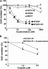
Figure 1
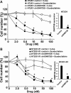
Figure 1. EC cells are highly sensitive to low concentrations of guadecitabine
(A) Cisplatin sensitive EC cells, NT2/D1, and cisplatin resistant cells, NT2/D1-R1, but not HCT116 colon cancer cells, U2OS osteosarcoma cells, or MCF7 breast cancer cells are sensitive to low concentrations of guadecitabine. Guadecitabine was added for 3 days to exponentially growing cultures. Viable cell growth and survival were measured. All data points are the average of biological triplicates. Error bars are standard deviation. *p < 0.01 comparing drug treatments to vehicle control in the same cell line. (B) Pretreatment with low concentrations of guadecitabine restores cisplatin sensitivity to cisplatin resistant EC cells. NT2/D1-R1 cells were pretreated with vehicle or 10 nM guadecitabine for 3 days before replating and a 48-hour recovery period followed by indicated cisplatin treatments for 6 hours. Cell viability was measured 3 days later. All data points are the average of biological triplicates. Error bars are standard deviation. *p < 0.01 comparing NTD1-R1 to NT2D1-R1 + guadecitabine. Experiments were repeated twice with similar results.
Figure 2
Figure 2. Guadecitabine and 5-aza sensitivity in EC cells are dependent on DNMT3B
DNMT3B knockdown results in resistance to guadecitabine and 5-aza in (A) NT2/D1 and (B) NT2/D1-R1 cells. Guadecitabine or 5-aza was added for 3 days to exponentially growing cultures. Viable cell growth and survival were measured. All data points are the average of biological triplicates. Error bars are standard deviation. *p < 0.01 and **p < 0.05 comparing control cell lines with their corresponding shDNMT3B cells. Right, The shDNMT3B cells were previously characterized and have a knockdown at the protein level of greater than 90% (18) and knockdown was further confirmed by real-time PCR assays. Error bars are standard deviation. *p < 0.01 comparing control cell lines with their corresponding shDNMT3B cells. Experiments were repeated twice with similar results.
Figure 3
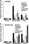
Figure 3. Guadecitabine and 5-aza remodel gene transcription in EC cells
(A) NT2/D1 and (B) NT2/D1-R1 cells were treated with 10 nM 5-aza or guadecitabine for 3 days and RNA was harvested for real-time PCR analysis. Each bar represents the average of 3 biological replicates. Error bars are standard deviation. *p < 0.01 and **p < 0.05 comparing each drug treatment to vehicle treatment. Experiments were repeated twice with similar results.
Figure 4
Figure 4. Guadecitabine potently inhibits growth and induces regression of cisplatin resistant EC tumors in vivo
(A) Mice bearing cisplatin resistant tumors were randomized to subcutaneous injections of guadecitabine at a dose of 2.0 mg/kg or vehicle control. Randomization and injections were performed when palpable tumors were just evident. Treatments were for 5 days per week for 2 weeks. Each line represents a single mouse (7 vehicle treated, 7 guadecitabine treated). (B) Representative tumors from the experiment in A. In most cases, tumors completely regressed after guadecitabine therapy while in a few cases a residual remnant tumor remained. (C) Treatment of 2.0 mg/kg guadecitabine for 5 days per week for 2 weeks did not effect whole body weight. Measurements began on the day after the first guadecitabine injection. (D) Mice bearing cisplatin resistant tumors were randomized to vehicle control or indicated dosages of guadecitabine for 5 days per week for 2 weeks. Each line represents a single mouse (3 mice per group). Note: some tumors were larger than just palpable size at the start of therapy and still regressed completely with guadecitabine; however, one mouse at the lowest, 0.1 mg/kg dose had only a partial response. (E) Low dose guadecitabine and cisplatin combination therapy is effective for cisplatin resistant EC cells. Mice bearing cisplatin resistant tumors were randomized to 3 groups. Guadecitabine alone at a dose of 0.5 mg/kg for 5 days for only 1 week, guadecitabine at a dose of 0.5 mg/kg for 5 days followed two days later with a single dose of 9 mg/kg cisplatin, or two doses of 9 mg/kg cisplatin on consecutive days. Each line represents a single mouse (4 mice per group). Note that two-dose cisplatin treatment alone resulted in lethal toxicity in two mice on day 12. No overt toxicity was seen in guadecitabine or guadecitabine + single dose cisplatin groups.
Figure 5
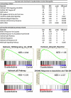
Figure 5. Guadecitabine induces p53 and immune pathway gene expression signatures in EC tumors in vivo
GSEA analysis of tumors treated in vivo for 4 days with 1.5 mg/kg guadecitabine (SGI-110) in biological triplicate revealed that the top gene sets enriched in response to guadecitabine are related to immune or p53 pathway activation. NES is Normalized Enrichment Score.
Figure 6
Figure 6. Guadecitabine induces HLA, NFKB and cancer testis antigens in EC tumors
(A) Microarray results for HLA class I and NFKB pathway genes. (B) Real-time PCR analysis of cancer testis antigens IMAGE-A3, IMAGE-A1 and NFKB gene, NFKBIA.
Figure 7
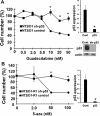
Figure 7. Guadecitabine and 5-aza sensitivity in EC cells is dependent on p53
Knockdown of p53 results in resistance to (A) Guadecitabine in NT2/D1 cells and (B) 5-aza in NT2/D1-R1 cells. Guadecitabine or 5-aza was added for 3 days to exponentially growing cultures. Viable cell growth and survival were measured. All data points are the average of biological triplicates. Error bars are standard deviation. *p < 0.01 comparing control cell lines with their corresponding shp53 cells. Right, The shp53 knockdown was confirmed by real-time PCR assays. Error bars are standard deviation. *p < 0.01 comparing control cell lines with their corresponding shp53 cells. Western blot showing robust knockdown of p53 in sh-p53 NT2/D1 cells is also included. A similar level of knockdown was also seen in NT2/D1-R1 cells (not shown). Experiments were repeated with similar results.

References

    1. Manecksha RP, Fitzpatrick JM. Epidemiology of testicular cancer. BJU Int. 2009;104:1329–1333. - PubMed
    1. Hanna NH, Einhorn LH. Testicular cancer—discoveries and updates. N Engl J Med. 2014;371:2005–2016. - PubMed
    1. Giuliano CJ, Freemantle SJ, Spinella MJ. Testicular germ cell tumors: a paradigm for the successful treatment of solid tumor stem cells. Curr Cancer Ther Rev. 2006;2:255–270. - PMC - PubMed
    1. Feldman DR, Patil S, Trinos MJ, Carousso M, Ginsberg MS, Sheinfeld J, Bajorin DF, Bosl GJ, Motzer RJ. Progression-free and overall survival in patients with relapsed/refractory germ cell tumors treated with single-agent chemotherapy: endpoints for clinical trial design. Cancer. 2012;118:981–986. - PubMed
    1. Porcu P, Bhatia S, Sharma M, Einhorn LH. Results of treatment after relapse from high-dose chemotherapy in germ cell tumors. J Clin Oncol. 2000;18:1181–1186. - PubMed

MeSH terms

Supplementary concepts