. 2017 Apr;188(1):22-35.
doi: 10.1111/cei.12912. Epub 2017 Jan 31.

T-bet over-expression regulates aryl hydrocarbon receptor-mediated T helper type 17 differentiation through an interferon (IFN)γ-independent pathway

Affiliations

T-bet over-expression regulates aryl hydrocarbon receptor-mediated T helper type 17 differentiation through an interferon (IFN)γ-independent pathway

M Yokosawa et al. Clin Exp Immunol. 2017 Apr.

Abstract

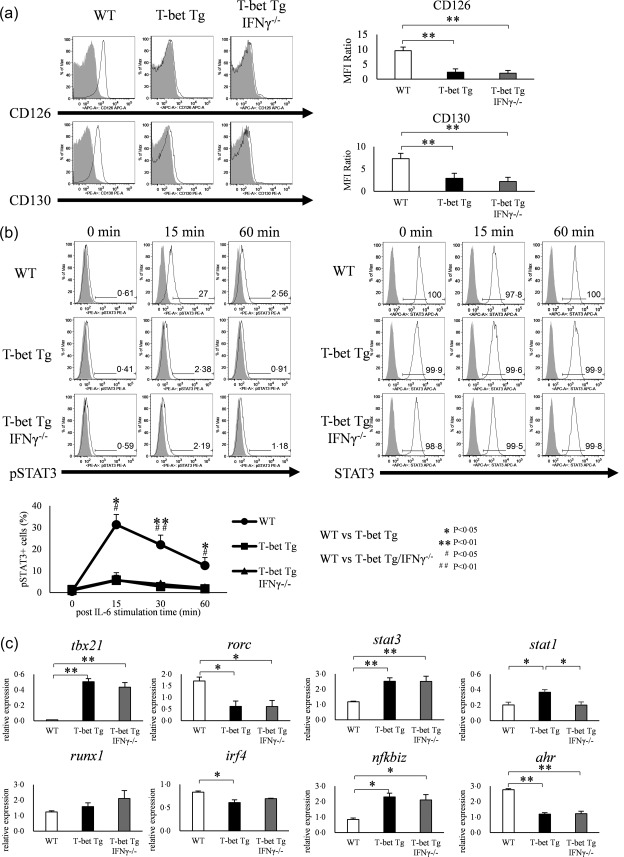
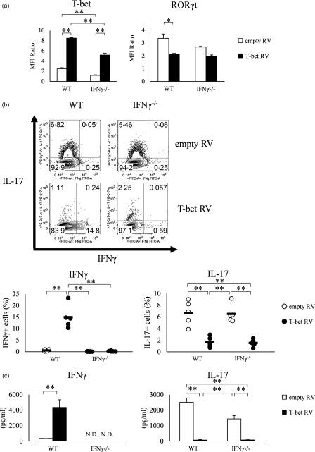
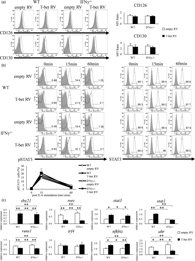
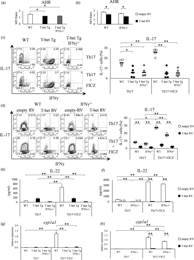
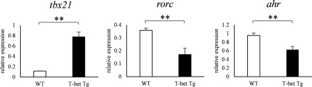
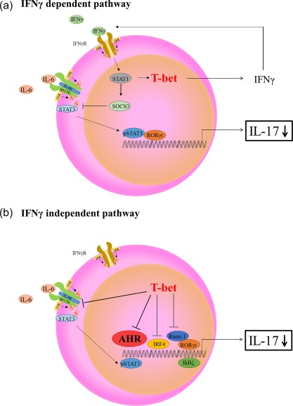
Various transcription factors are also known to enhance or suppress T helper type 17 (Th17) differentiation. We have shown previously that the development of collagen-induced arthritis was suppressed in T-bet transgenic (T-bet Tg) mice, and T-bet seemed to suppress Th17 differentiation through an interferon (IFN)-γ-independent pathway, although the precise mechanism remains to be clarified. The present study was designed to investigate further the mechanisms involved in the regulation of Th17 differentiation by T-bet over-expression, and we found the new relationship between T-bet and aryl hydrocarbon receptor (AHR). Both T-bet Tg mice and IFN-γ-/- -over-expressing T-bet (T-bet Tg/IFN-γ-/- ) mice showed inhibition of retinoic acid-related orphan receptor (ROR)γt expression and IL-17 production by CD4+ T cells cultured under conditions that promote Th-17 differentiation, and decreased IL-6 receptor (IL-6R) expression and signal transducer and activator of transcription-3 (STAT-3) phosphorylation in CD4+ T cells. The mRNA expression of ahr and rorc were suppressed in CD4+ T cells cultured under Th-17 conditions from T-bet Tg mice and T-bet Tg/IFN-γ-/- mice. CD4+ T cells of wild-type (WT) and IFN-γ-/- mice transduced with T-bet-expressing retrovirus also showed inhibition of IL-17 production, whereas T-bet transduction had no effect on IL-6R expression and STAT-3 phosphorylation. Interestingly, the mRNA expression of ahr and rorc were suppressed in CD4+ T cells with T-bet transduction cultured under Th17 conditions. The enhancement of interleukin (IL)-17 production from CD4+ T cells by the addition of AHR ligand with Th17 conditions was cancelled by T-bet over-expression. Our findings suggest that T-bet over-expression-induced suppression of Th17 differentiation is mediated through IFN-γ-independent AHR suppression.

Keywords: AHR; IL-17; T-bet over-expression.


Figures

Figure 1
Figure 1
Over‐expression of T‐bet in transgenic (Tg) mice suppresses IL‐17 expression independently of interferon (IFN)‐γ. (a) IFN‐γ and interleukin (IL)−17 production by splenic CD4+ T cells of wild‐type (WT), T‐bet Tg and T‐bet Tg/IFN‐γ–/– mice CD4+ analysed by flow cytometry. Numbers in plots indicate percentage of cells that secrete the cytokines. Five mice per group were analysed under the T helper type 17 (Th17) condition and representative data are shown with neutral condition. (b) T‐bet and retinoic acid‐related orphan receptor (RORγt) expression on CD4+ cells analysed by flow cytometry. Five mice per group were analysed under the Th17 condition. Data are mean ± standard error of the mean (s.e.m.) of four mice. *P < 0·05; ** P < 0·01 (by Tukey's test).
Figure 2
Figure 2
Over‐expression of T‐bet in transgenic (Tg) mice modulates the interleukin (IL)−6/signal transducer and activation of transcription‐3 (STAT‐3) pathway and regulates the expression and function of other transcription factors. (a) Expression of CD126 (IL‐6Rα) and CD130 (IL‐6Rβ) on CD4+ T cells of wild‐type (WT), T‐bet Tg and T‐bet Tg/interferon (IFN)‐γ–/– mice analysed by flow cytometry (open histogram; CD126 or CD130, shaded histogram; isotype control). Five mice per group were analysed and representative data are shown. *P < 0·05; **P < 0·01 (by Tukey's test). (b) STAT‐3 phosphorylation analyzed by flow cytometry in splenic CD4+ T cells of WT, T‐bet Tg and T‐bet Tg/IFN‐γ–/– mice stimulated with IL‐6 for 0, 15, 30 and 60 min (open histogram; phosphorylated STAT‐3 or STAT‐3, shaded histogram; isotype control or blank). Five mice per group were analysed and representative data are shown. *P < 0·05; **P < 0·01 (by Tukey's test and Kruskal–Wallis test). (c) Expression of tbx21, rorc, stat3, stat1, runx1, irf4, nfkbiz and ahr in CD4+ T cells of WT, T‐bet Tg and T‐bet Tg/IFN‐γ–/– mice analysed by quantitative reverse transcription–polymerase chain reaction (qRT–PCR). Four mice per group were analysed. Data are mean ± standard error of the mean (s.e.m.) of four mice. *P< 0·05; **P < 0·01 (by Tukey's test).
Figure 3
Figure 3
Over‐expression of T‐bet by retroviral transduction into naive CD4+ T cells of wild‐type (WT) and interferon (IFN)‐γ–/– mice suppresses retinoic acid‐related orphan receptor (RORγt) expression and interleukin (IL)−17 production. (a) T‐bet and RORγt expression in retroviral transfected splenic CD4+ T cells of WT and IFN‐γ–/– mice, analysed by flow cytometry. Data of five experiments were analysed. (b) IFN‐γ and IL‐17 production analysed by flow cytometry. Data of five experiments were analysed and representative data are shown. Numbers in plots indicate percentage of cells that secrete the indicated cytokines. (c) IFN‐γ and IL‐17 production in the culture supernatant, as determined by enzyme‐linked immunosorbent assay (ELISA). Data are mean ± standard error of the mean (s.e.m.) of four experiments; n.d. = not detected. *P < 0·05; **P < 0·01 (by Tukey's test).
Figure 4
Figure 4
Over‐expression of T‐bet in CD4+ T cells neither suppresses the expression of interleukin (IL)−6R nor phosphorylation of signal transducer and activation of transcription‐3 (STAT‐3). (a) Expression of CD126 (IL‐6Rα) and CD130 (IL‐6Rβ) on CD4+ T cells of wild‐type (WT) and interferon (IFN)‐γ–/– mice transfected with retroviral vector (open histogram; CD126 or CD130, shaded histogram; isotype control). Data are mean ± standard error of the mean (s.e.m.) of four experiments. (b) signal transducer and activation of transcription‐3 (STAT3) phosphorylation in CD4+ T cells of WT and IFN‐γ–/– mice transfected with retroviral vector and stimulated with IL‐6 for 0, 15 and 60 min, as analysed by flow cytometry (open histogram; phosphorylated STAT‐3 or STAT‐3, shaded histogram; isotype control or blank). Three experiments were analysed and representative data are shown. Data are mean ± s.e.m. of three experiments. *P < 0·05; **P < 0·01 (by Tukey's test). (c) CD4+ T cells of WT and IFN‐γ–/– mice transfected with retroviral vector and analysed for the expression of tbx21, rorc, stat3, stat1, runx1, irf4, nfkbiz and ahr by quantitative reverse transcription–polymerase chain reaction (qRT–PCR). Data are mean ± s.e.m. of five experiments. *P < 0·05; **P < 0·01 (by Tukey's test).
Figure 5
Figure 5
T‐bet over‐expression suppresses aryl hydrocarbon receptor (AHR) expression independent of interferon (IFN)‐γ. (a) CD4+ T cells of wild‐type (WT), T‐bet transgenic (Tg) and T‐bet Tg/IFN‐γ–/– mice analysed for the AHR expression by flow cytometry. Four mice per group were analysed. (b) CD4+ T cells of WT and IFN‐γ–/– mice transfected with retroviral vector and analysed for AHR expression by flow cytometry. Four experiments per group were analysed. (c) CD4+ T cells of WT, T‐bet Tg and T‐bet Tg/IFN‐γ–/– mice cultured with or without 6‐formylindolo (3,2‐b) carbazole (FICZ) and analysed for IFN‐γ and interleukin (IL)−17 production by flow cytometry. Five mice per group were analysed and representative data are shown. (d) CD4+ T cells of WT and IFN‐γ–/– mice transfected with retroviral vector cultured with or without FICZ, and analysed for IFN‐γ and IL‐17 production by flow cytometry. Three experiments per group were analysed and representative data are shown. (e) The production of IL‐22 analysed by enzyme‐linked immunosorbent assay (ELISA) in the experiment described in (c). (f) The production of IL‐22 analysed by ELISA in the experiment described in (d). (g) Expression levels of cyp1a1 analysed by quantitative reverse transcription–polymerase chain reaction (qRT–PCR) in the experiment described in (c). (h) Expression level of cyp1a1 analysed by qRT–PCR in the experiment described in (d). *P < 0·05; **P < 0·01 (by Tukey's test).
Figure 6
Figure 6
The mRNA expression of ahr was suppressed in collagen‐induced arthritis (model) of T‐bet transgenic (Tg) mice. Ten days after the collagen type II (CII) immunization, lymph node cells from wild‐type (WT) and T‐bet Tg mice were cultured for 72 h in the presence of 100 μg/ml of denatured CII. After culture of lymph node cells with CII, CD4+ cells were purified by magnetic cell isolation and cell separation (MACS), and cDNA was obtained. The levels of tbx21, rorc and ahr expression were analysed by reverse transcription–polymerase chain reaction (RT–PCR). Data are mean ± standard error of the mean (s.e.m.) of three mice. **P < 0·01 (by Student's t‐test).
Figure 7
Figure 7
Schematic diagram of potential mechanisms involved in T helper type 17 (Th17) differentiation under T‐bet over‐expression conditions. (a) Interferon (IFN)‐γ‐dependent T‐bet suppression of Th17 differentiation. IFN‐γ signal transducer and activation of transcription‐1 (STAT‐1) signalling induces suppressor of cytokine signaling 3 (SOCS3) expression, inhibition of IL‐6/STAT‐3 and retinoic acid‐related orphan receptor (RORγt) expression, resulting in suppression of interleukin (IL)−17 production. (b) IFN‐γ‐independent T‐bet suppression of Th‐17 differentiation. T‐bet over‐expression regulates various transcription factors [RORγt, runt‐related transcription factor 1 (Runx‐1), I kappa B zeta (IκBζ), IFN regulatory factor 4 (IRF4) and aryl hydrocarbon receptor (AHR)], resulting in suppression of IL‐17 production. Our findings particularly support the possibility that the regulatory mechanism of Th17 differentiation might be due to the suppression of AHR.

References

    1. Jetten AM. Retinoid‐related orphan receptors (RORs): critical roles in development, immunity, circadian rhythm, and cellular metabolism. Nucl Recept Signal 2009; 7:e003. - PMC - PubMed
    1. Tesmer LA, Lundy SK, Sarkar S, Fox DA. Th17 cells in human disease. Immunol Rev 2008; 223:87–113. - PMC - PubMed
    1. Szabo SJ, Kim ST, Costa GL, Zhang X, Fathman CG, Glimcher LH. A novel transcription factor, T‐bet, directs Th1 lineage commitment. Cell 2000; 100:655–69. - PubMed
    1. Szabo SJ, Sullivan BM, Stemmann C, Satoskar AR, Sleckman BP, Glimcher LH. Distinct effects of T‐bet in TH1 lineage commitment and IFN‐γ production in CD4 and CD8 T cells. Science 2002; 295:338–42. - PubMed
    1. Afkarian M, Sedy JR, Yang J et al T‐bet is a STAT1‐induced regulator of IL‐12R expression in naïve CD4+ T cells. Nat Immunol 2002; 3:549–57. - PubMed

Publication types

MeSH terms