

Quantitative radiomics studies for tissue characterization: a review of technology and methodological procedures
- PMID: 27936886
- PMCID: PMC5685111
- DOI: 10.1259/bjr.20160665
Quantitative radiomics studies for tissue characterization: a review of technology and methodological procedures
Abstract
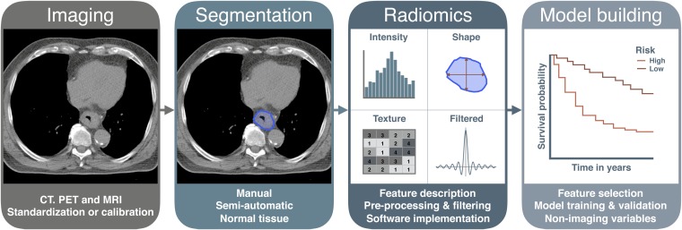
Quantitative analysis of tumour characteristics based on medical imaging is an emerging field of research. In recent years, quantitative imaging features derived from CT, positron emission tomography and MR scans were shown to be of added value in the prediction of outcome parameters in oncology, in what is called the radiomics field. However, results might be difficult to compare owing to a lack of standardized methodologies to conduct quantitative image analyses. In this review, we aim to present an overview of the current challenges, technical routines and protocols that are involved in quantitative imaging studies. The first issue that should be overcome is the dependency of several features on the scan acquisition and image reconstruction parameters. Adopting consistent methods in the subsequent target segmentation step is evenly crucial. To further establish robust quantitative image analyses, standardization or at least calibration of imaging features based on different feature extraction settings is required, especially for texture- and filter-based features. Several open-source and commercial software packages to perform feature extraction are currently available, all with slightly different functionalities, which makes benchmarking quite challenging. The number of imaging features calculated is typically larger than the number of patients studied, which emphasizes the importance of proper feature selection and prediction model-building routines to prevent overfitting. Even though many of these challenges still need to be addressed before quantitative imaging can be brought into daily clinical practice, radiomics is expected to be a critical component for the integration of image-derived information to personalize treatment in the future.
Figures
References
-
- Eisenhauer EA, Therasse P, Bogaerts J, Schwartz LH, Sargent D, Ford R, et al. . New response evaluation criteria in solid tumours: revised RECIST guideline (version 1.1). Eur J Cancer 2009; 45: 228–47. doi: https://doi.org/10.1016/j.ejca.2008.10.026 - DOI - PubMed
-
- Wahl RL, Jacene H, Kasamon Y, Lodge MA. From RECIST to PERCIST: evolving considerations for PET response criteria in solid tumors. J Nucl Med 2009; 50(Suppl 1): 122S–50S. doi: https://doi.org/10.2967/jnumed.108.057307 - DOI - PMC - PubMed
-
- de Bruijne M. Machine learning approaches in medical image analysis: from detection to diagnosis. Med Image Anal 2016; 33: 94–7. doi: https://doi.org/10.1016/j.media.2016.06.032 - DOI - PubMed
-
- Abe Y, Hanai K, Nakano M, Ohkubo Y, Hasizume T, Kakizaki T, et al. . A computer-aided diagnosis (CAD) system in lung cancer screening with computed tomography. Anticancer Res 2005; 25: 483–8. - PubMed
-
- Li F. Potential clinical impact of advanced imaging and computer-aided diagnosis in chest radiology: importance of radiologist's role and successful observer study. Radiol Phys Technol 2015; 8: 161–73. doi: https://doi.org/10.1007/s12194-015-0319-0 - DOI - PubMed
Publication types
MeSH terms
LinkOut - more resources
Full Text Sources
Other Literature Sources
Medical
