

Review
doi: 10.1259/bjr.20160607.
Epub 2016 Dec 12.
Hamartomas from head to toe: an imaging overview
Affiliations
- PMID: 27936889
- PMCID: PMC5601532
- DOI: 10.1259/bjr.20160607
Item in Clipboard
Review
Hamartomas from head to toe: an imaging overview
Br J Radiol.
2017 Mar.
Abstract
Hamartomas are tumours composed of mesenchymal tissues such as cartilage, fat, connective tissue and smooth muscle and can be found in virtually any organ system. These masses commonly develop sporadically, but are also seen in certain syndromes such as tuberous sclerosis or Carney triad. While their imaging appearance varies depending on the organ they arise from, findings are usually unique and a diagnosis can be confidently made. Radiologists must be aware of the clinical and imaging presentations of these lesions with the particular goal of avoiding unnecessary studies or invasive procedures. Furthermore, knowledge of common syndromic entities is crucial, as the radiologist may be the first to suggest the diagnosis.
Figures

Tuber cinereum hamartoma: an axial T2 weighted MR image of the brain is showing an isointense mass (arrow) posterior to the pituitary stalk compatible with a tuber cinereum hamartoma.

Cortical tubers in a 28-year-old male with tuberous sclerosis: an axial fluid-attenuated inversion-recovery MR image of the brain is demonstrating multiple areas of hyperintense signal with associated gyral expansion in the bilateral frontal and parietal lobes (arrows).

Multiple subependymal nodules in a patient with tuberous sclerosis: an axial T2 weighted MR image of the brain at the level of the lateral ventricles is showing multiple isointense to hypointense subependymal nodules bilaterally (arrows). The hypointense signal is in keeping with calcification.
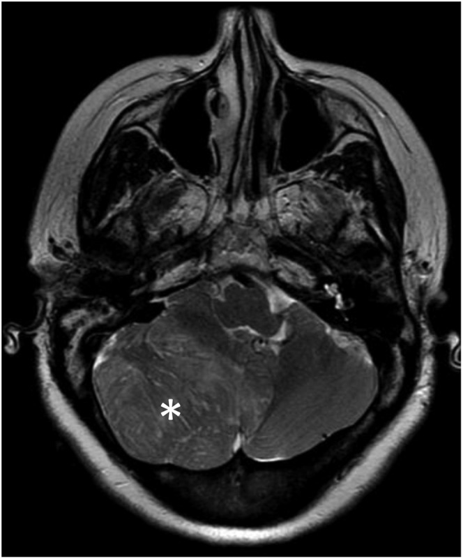
Cerebellar hamartoma (Lhermitte–Duclos disease): the axial T2 weighted MR of the brain at the level of the cerebellum is demonstrating a large, hyperintense, intra-axial mass replacing most of the right cerebellar hemisphere (asterisk), causing a moderate mass effect over the fourth ventricle and pons. The thickened gyri and bizarre “gyriform” appearance can be noted.

Nasal chondromesenchymal hamartoma: an axial CT image (a) is showing a mildly lobulated, partially calcified soft-tissue mass (arrow) within the right nasal cavity, producing a mass effect and bony remodelling without frank bone destruction. A coronal T2 weighted (b) MR image is showing a heterogeneous, slightly hyperintense mass (arrow).

Pulmonary hamartoma: a contrast-enhanced CT image through the lower lobes is showing a large, lobulated soft-tissue density mass in the left lower lobe with foci of low attenuation and calcification.

Multifocal micronodular pneumocyte hyperplasia in a patient with tuberous sclerosis: CT of the chest on lung window is showing multiple subcentimetre nodules (arrows).

Cardiac rhabdomyoma: a T2 weighted dark-blood axial MR image through the heart is shows a large well-defined mass arising off the anterolateral wall of the left ventricle (arrow). The mass is appearing slightly hyperintense with respect to the myocardium.

Breast hamartoma: a craniocaudal mammogram of the left breast is demonstrating a large fat-containing mass in the medial aspect of the breast (arrows). The mass is showing a thin radiopaque capsule.

Hepatic mesenchymal hamartoma in a young infant: the axial T2 weighted (a) and post-contrast T1 weighted (b) MR images of the abdomen are demonstrating a large multicystic mass arising from the liver. No enhancing nodules are seen. Case courtesy of Christine Menias, MD.

Biliary hamartomas (von Meyenburg complex): a contrast-enhanced CT image of the abdomen is demonstrating innumerable fluid-attenuating lesions throughout the hepatic parenchyma. There are no internal septations or nodularity. Case courtesy of Srinivasa Prasad, MD.

Multiple bilateral renal angiomyolipomas (AMLs): a transaxial contrast-enhanced CT image of the abdomen is showing multiple heterogeneous lesions arising from the bilateral kidneys (black arrows), many of which are demonstrating foci of fat attenuation. A predominantly fatty lesion is seen in the right kidney (white arrowhead). Arising from the anterior aspect of the right renal interpolar region is an enhancing lesion that is predominantly homogeneous and compatible with a lipid-poor renal AML (white arrow).

Fibrolipomatous hamartoma of the sciatic nerve: an axial T1 weighted MR image through the left gluteal region is demonstrating increased thickness and fatty infiltration of the sciatic nerve (arrow), resulting in a “coaxial cable” appearance.

Gastrointestinal stromal tumour (GIST) and pulmonary chondroma in a patient with Carney triad: the contrast-enhanced CT of the abdomen (a) is showing a well-delineated endophytic soft-tissue density mass extruding into the gastric lumen in the region of the stomach fundus (arrow). The patient presented two other similar masses (not shown). CT of the chest in the same patient (b) is demonstrating a large, lobulated left upper lobe mass with coarse foci of calcification. In the setting of multiple GISTs, and given the CT appearance of the pulmonary lesion, this is compatible with a pulmonary chondroma.

Lymphangioleiomyomatosis (LAM) in a patient with tuberous sclerosis: a high-resolution axial CT image of the chest is demonstrating innumerable somewhat uniform cysts throughout the lung parenchyma, with findings compatible with TSC-associated LAM.

Proteus syndrome in a in a young female: a photograph of the patient hands (a) is showing an abnormal asymmetric growth of the left index finger and to a lesser degree of the distal aspect of the left middle finger. The frontal radiograph of the left hand (b) is showing an asymmetric growth of the left index finger phalanges and thickening of the overlying soft tissues.
References
-
- Albrecht E. Über hamartome. Verh Dtsch Ges Pathol 1904; 7: 153–7.
-
- Maitra A. Robbins and Cotran pathologic basis of disease. 9th edn. Amsterdam, Netherlands: Elsevier; 2015.
-
- Saleem SN, Said AH, Lee DH. Lesions of the hypothalamus: MR imaging diagnostic features. Radiographics 2007; 27: 1087–108. doi: https://doi.org/10.1148/rg.274065123 - DOI - PubMed
-
- Glastonbury CM, Osborn AG, Salzman KL. Masses and malformations of the third ventricle: normal anatomic relationships and differential diagnoses. Radiographics 2011; 31: 1889–905. doi: https://doi.org/10.1148/rg.317115083 - DOI - PubMed
Publication types
MeSH terms
LinkOut - more resources
Full Text Sources
Other Literature Sources
Medical
