Review
. 2017 Feb 10;17(2):141-154.
doi: 10.3727/105221616X693639. Epub 2016 Nov 2.

Novel Advances in Understanding of Molecular Pathogenesis of Hepatoblastoma: A Wnt/β-Catenin Perspective

Review

Novel Advances in Understanding of Molecular Pathogenesis of Hepatoblastoma: A Wnt/β-Catenin Perspective

Danielle Bell et al. Gene Expr. .

Abstract

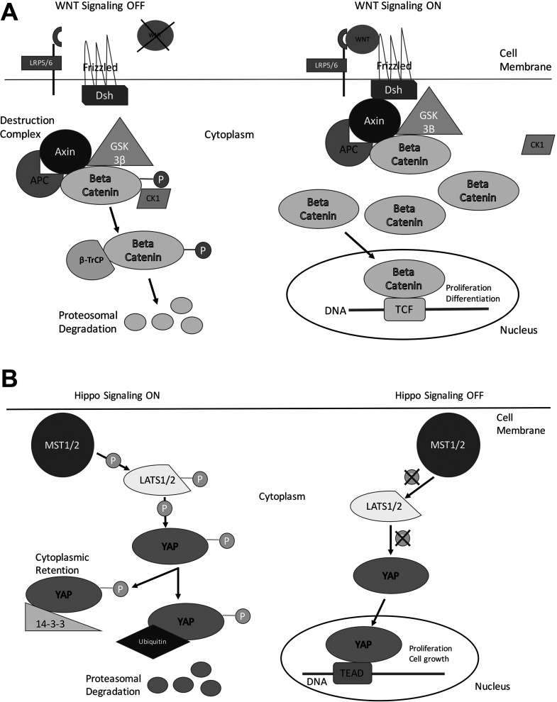
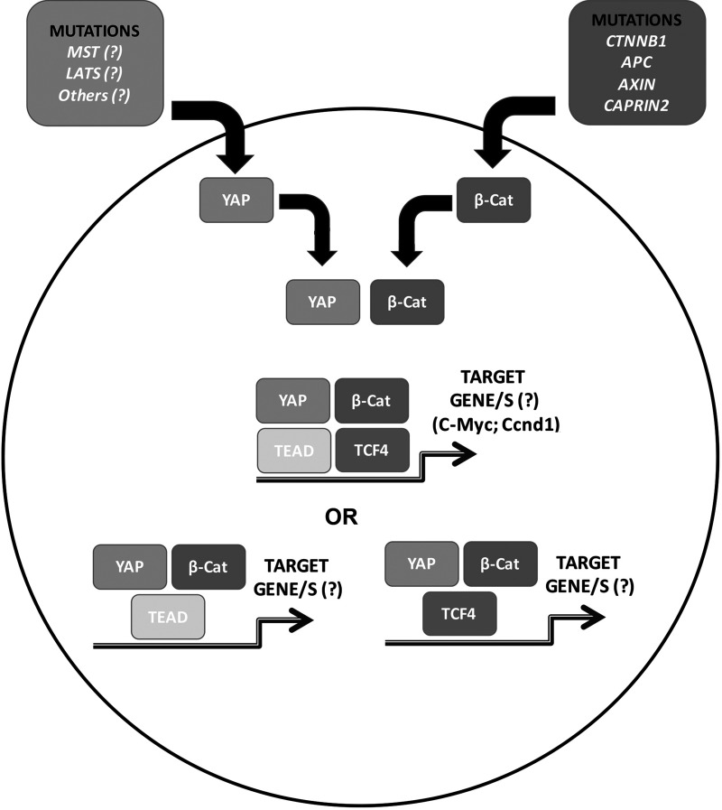
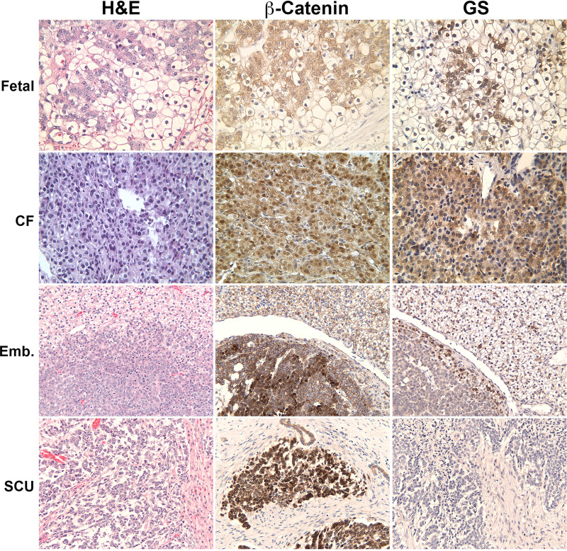
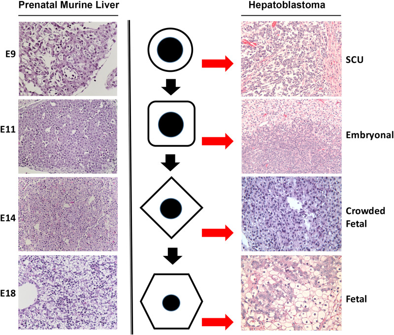
Hepatoblastoma is the most common pediatric liver malignancy, typically striking children within the first 3 years of their young lives. While advances in chemotherapy and newer surgical techniques have improved survival in patients with localized disease, unfortunately, for the 25% of patients with metastasis, the overall survival remains poor. These tumors, which are thought to arise from hepatic progenitors or hepatoblasts, hence the name hepatoblastoma, can be categorized by histological subtyping based on their level of cell differentiation. Genomic and histological analysis of human tumor samples has shown exon-3 deletions or missense mutations in gene coding for β-catenin, a downstream effector of the Wnt signaling pathway, in up to 90% of hepatoblastoma cases. The current article will review key aberrations in molecular pathways that are implicated in various subtypes of hepatoblastoma with an emphasis on Wnt signaling. It will also discuss cooperation among components of pathways such as β-catenin and Yes-associated protein in cancer development. Understanding the complex network of molecular signaling in oncogenesis will undoubtedly aid in the discovery of new therapeutics to help combat hepatoblastoma.


Figures

Figure 1
Figure 1
Simplified overview of the Wnt/β-catenin signaling and the Hippo signaling pathways. (A) In “off” state, β-catenin complexes with the members of its degradation complex where it is phosphorylated at specific serine and threonine residues for recognition by proteasome for degradation. In its “on” state, Wnt binds to receptor Frizzled and coreceptor LRP5/6 to recruit dishevelled, allowing for the β-catenin degradation complex to be inactivated. This leads to hypophosphorylation of β-catenin, its stabilization in the cytoplasm followed by its nuclear translocation. In the nucleus, β-catenin acts as a cofactor for TCF family of transcription factors to induce target gene expression. (B) In its “on” state, the Hippo signaling kinases MST1/2 are activated to in turn activate the LATS1/2 kinases. The activated LATS1/2 in turn phosphorylates YAP (or its paralog TAZ), the major effector of the Hippo cascade. Once phosphorylated, YAP (or TAZ) is either retained in the cytoplasm or degraded by proteasome. In its “off” state, the kinase complexes in the Hippo signaling are inactive, and YAP (or TAZ) can translocate to the nucleus to act as cofactor for the TEAD family of transcription factors and regulate expression of key target genes.
Figure 2
Figure 2
Yap and β-catenin coexpression in liver leads to hepatoblastomas in mice. Gross image of tumor-bearing liver at 9 weeks after hydrodynamic tail vein injection of sleeping beauty plasmids carrying mutant Yap and β-catenin. Histology from a representative 9-week postinjection liver reveals the tumors to be hepatoblastomas. Tumors were notably positive for nuclear and cytoplasmic β-catenin and strongly positive for the β-catenin target cyclin D1. Tumors were negative for another β-catenin target, glutamine synthetase (GS). Tumors were strongly positive for β-catenin target cyclin D1.
Figure 3
Figure 3
Model depicting Yap and β-catenin cooperation in hepatoblastoma. Nuclear translocation of β-catenin in the majority of hepatoblastomas occurs due to various mechanisms such as mutations in CTNNB1 or due to mutations affecting genes encoding for proteins like Axin1 and APC, which are required for β-catenin degradation. Yap nuclear translocation also is evident in a majority of these tumors; however, the mechanism remains elusive. In the nucleus, the two proteins interact with each other and either interact their respective transcription factors together in a complex or in two separate complexes as indicated to eventually dictate target genes such as cyclin D1 and c-Myc or others that are not yet identified, to lead to hepatoblastoma development and growth.
Figure 4
Figure 4
Wnt signaling status in various histologic subtypes of hepatoblastoma in patients. A representative fetal hepatoblastoma shows the presence of nuclear and cytoplasmic β-catenin along with patchy positivity for glutamine synthetase (GS). A crowded fetal (CF) hepatoblastoma is positive for β-catenin as well as GS, which is more homogeneous. Embryonal hepatoblastoma is strongly positive for nuclear and cytoplasmic β-catenin but negative for GS. Occasional area may show some staining for GS. Small cell undifferentiated (SCU) hepatoblastoma is again strongly positive for β-catenin but completely lacks any GS staining.
Figure 5
Figure 5
Comparative histology of murine prenatal hepatic developmental stages and histologic subtypes of human hepatoblastoma. Hepatoblastomas are classified based on their cell composition. There is resemblance in histology of various stages of hepatic development to various subtypes of hepatoblastoma, which is dictated by the predominant cell type that arrests and expands within the tumor. Shown to the left are representative images of developing mouse liver from embryonic day 9 (E9), E11, E14, and E18. Shown to the right are the sections from patient hepatoblastomas of various subtypes including small cell undifferentiated (SCU), embryonal (Emb), crowded fetal (CF), and fetal hepatoblastoma. The middle row is a representation of the predominant cell type that exists at specific stages during normal liver development, but an arrest at specific stages of maturation leads to their expansion and enrichment to eventually contribute to a specific histology and eventually a histological subtype of hepatoblastoma.

References

    1. Darbari A, Sabin KM, Shapiro CN, Schwarz KB. Epidemiology of primary hepatic malignancies in U.S. children. Hepatology 2003;38(3):560–6. - PubMed
    1. McLaughlin CC, Baptiste MS, Schymura MJ, Nasca PC, Zdeb MS. Maternal and infant birth characteristics and hepatoblastoma. Am J Epidemiol. 2006;163(9):818–28. - PubMed
    1. Czauderna P, Haeberle B, Hiyama E, Rangaswami A, Krailo M, Maibach R, Rinaldi E, Feng Y, Aronson D, Malogolowkin M, Yoshimura K, Leuschner I, Lopez-Terrada D, Hishiki T, Perilong G, von Schewinitz D, Schmid I, Watanabe K, Derosa M, Meyers R. The Children’s Hepatic tumors International Collaboration (CHIC): Novel global rare tumor database yields new prognostic factors in hepatoblastoma and becomes a research model. Eur J Cancer 2016;52:92–101. - PMC - PubMed
    1. Tanimura M, Matsui I, Abe J, Ikeda H, Kobayashi N, Ohira M, Yokoyama M, Kaneko M. Increased risk of hepatoblastoma among immature children with a lower birth weight. Cancer Res. 1998;58(14):3032–5. - PubMed
    1. Spector LG, Johnson KJ, Soler JT, Puumala SE. Perinatal risk factors for hepatoblastoma. Br J Cancer 2008;98(9):1570–3. - PMC - PubMed

MeSH terms