Review
. 2016 Dec:18:1-10.
doi: 10.1016/j.cois.2016.08.001. Epub 2016 Aug 16.

Neuronal migration during development and the amyloid precursor protein

Affiliations
Review

Neuronal migration during development and the amyloid precursor protein

Philip F Copenhaver et al. Curr Opin Insect Sci. 2016 Dec.

Abstract

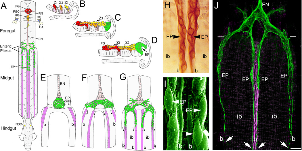
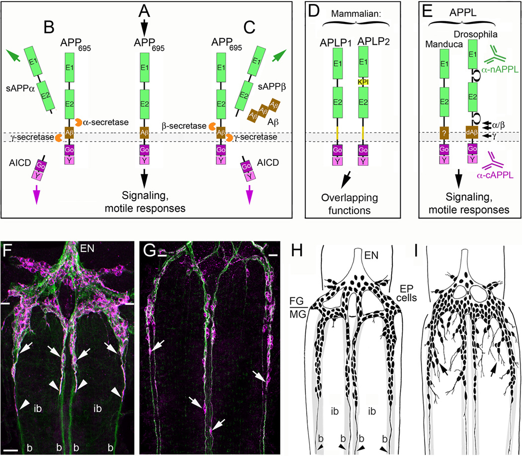
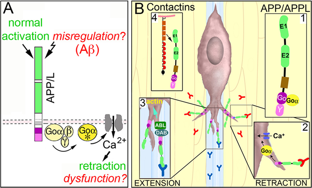
The Amyloid Precursor Protein (APP) is the source of amyloid peptides that accumulate in Alzheimer's disease. However, members of the APP family are strongly expressed in the developing nervous systems of invertebrates and vertebrates, where they regulate neuronal guidance, synaptic remodeling, and injury responses. In contrast to mammals, insects express only one APP ortholog (APPL), simplifying investigations into its normal functions. Recent studies have shown that APPL regulates neuronal migration in the developing insect nervous system, analogous to the roles ascribed to APP family proteins in the mammalian cortex. The comparative simplicity of insect systems offers new opportunities for deciphering the signaling mechanisms by which this enigmatic class of proteins contributes to the formation and function of the nervous system.


Figures

Figure 1
Figure 1. Embryonic development of the insect Enteric Nervous System (ENS) involves extensive patterns of neuronal and glial migration
(A), Schematic drawing of the ENS and associated neurosecretory organs in the larval stage of the tobacco hornworm Manduca sexta (modified from [27]). The primary ganglion on the foregut is the frontal ganglion (FG; red), connected to the overlying brain lobes by paired frontal ganglion connectives (FGC). Several nerve branches extend anteriorly onto the pharynx, while the recurrent nerve (RN) extends posteriorly to the hypocerebral ganglion (HG; orange), situated below the brain. In Manduca, the hypocerebral ganglion initially forms during embryogenesis but then becomes closely opposed to the frontal ganglion and is no longer readily distinguished in later stages. The HG is also connected to the paired corpora cardiaca (CC; blue), the primary neurosecretory organs of the brain, which are adjacent to the corpora allata (CA; the source of Juvenile Hormone). From the HG, the esophageal nerve (EN) extends posteriorly along the length of the foregut, giving rise to short nerve branches that innervate the foregut musculature. Near the foregut-midgut boundary, the esophageal nerve connects with the enteric plexus that spans the foregut-midgut boundary, which includes nerve branches extending along radial muscles on the foregut and major nerves that extend along eight well-defined muscle bands that lie superficially on the midgut (purple). The enteric plexus contains a population of ~300 distributed neurons (EP cells; green), which includes intermingled groups of neurons expressing a variety of morphological and transmitter phenotypes. The EP cells occupy positions along the anterior 20% of the midgut, and extend long axons posteriorly along the muscle bands with sparse lateral branches that provide a diffuse innervation of the interband midgut musculature. The hindgut is innervated by branches of the proctodeal and rectal nerves that originate in the terminal abdominal ganglion of the ventral nerve cord. Branches of the proctodeal nerve also extend onto the posterior midgut and contain several peripheral neurosecretory cells (yellow). (B–C), Neurogenesis of the developing ENS in Manduca (after [31]). Panels show lateral views of the foregut midline; anterior is to the left, dorsal is to the top. When raised at 25°C, Manduca embryogenesis is complete in 100 hr (1 hr = 1 hour post-fertilization, or hpf). (B), By ~24 hpf, three neurogenic zones (Z1, Z2, & Z3) have formed in the dorsal foregut epithelium, which give rise to a series of mitotically active precursor cells via sequential delamination. Precursors giving rise to neurons typically divide only once (or occasionally twice) after delaminating, similar to midline precursors in the embryonic CNS. (C), By 28 hpf, streams of zone-derived cells have begun to migrate anteriorly along the foregut, while the remaining zone 3 cells delaminate as a group. The epithelium surrounding the original position of zone 3 subsequently differentiates into a distinct placode that will form the EP cells (green). (D), By 33 hpf, migrating zone cells have begun to form the frontal ganglion (FG), while the remaining zone 2 cells delaminate as a group. The EP cell placode has also begun to invaginate from the EP cell packet (described below). Zone 1 continues to generate cells until almost 40 hpf (not shown); late-emerging zone cells derived from all three zones tend to become glial precursors that remain mitotically active throughout much of embryogenesis and establish the glial sheath surrounding the foregut nerves and ganglia. (E–F), Formation of the midgut enteric plexus; panels show dorsal views of the developing ENS at the foregut-midgut boundary (after [88]). (E), By 40 hpf, the EP cells (green) have invaginated en mass from their neurogenic placode located within the posterior dorsal lip of the foregut (D, green). The neurons then commence a bilateral spreading phase of migration (arrows) that almost completely encircles the foregut, adjacent to the foregut-midgut boundary. Concurrently, subsets of longitudinal muscles on the midgut (magenta) begin to coalesce into eight well-defined bands as dorsal closure of the midgut proceeds. Anteriorly, the EP cell packet is in continuity with the developing esophageal nerve (EN), which contains populations of proliferating glial precursors (pink; derived from zone 3) that will subsequently ensheath the enteric plexus. (F), By 55 hpf, the EP cells have almost completely surrounded the foregut, and subsets of the neurons have aligned with each of the midgut muscle bands (only the dorsal four are shown). (G), By 58 hpf, subsets of EP cells have begun to migrate in a chain-like manner along the midgut muscle bands; smaller subsets also migrate onto radial muscles of the foregut (muscles not shown). Proliferating glial cells (pink) subsequently migrate along the pathways established by the neurons, thereby ensheathing the branches of the enteric plexus. (H), Magnified view of EP cell groups migrating on the mid-dorsal band pathways (at 58 hpf) of an embryo immunostained with an antibody recognizing all isoforms of the cell adhesion receptor Fasciclin II (Fas II). The migratory neurons and underlying muscle bands (b) express transmembrane Fas II (TM-Fas II), while the trailing glial cells express GPI-linked Fas II. The migratory EP cells and their processes remain primarily confined to their band pathways while avoiding the adjacent interband musculature (ib). (I), Scanning electron micrograph showing the migratory EP cells on the mid-dorsal band pathways (b) of an embryo at 65 hpf. (J), Lower magnified view of the developing ENS at 62 hpf, in an embryo that was immunostained with anti-TM-Fas II (green). TM-Fas II immunoreactivity in the mid-dorsal muscle bands is shown in magenta to better distinguish the EP cell processes (after [79]). At this stage, the EP cells have migrated ~200 µm and have begun to extend fasciculated axons (arrows) more posteriorly along the muscle bands (b). Throughout this developmental period, the EP cells avoid the adjacent interband regions (ib), extending terminal branches onto the lateral musculature only after migration and axogenesis is complete (~80 hpf). Scale = 20 µm in (H); 5 µm in (I); 60 µm in (J).
Figure 2
Figure 2. The insect ortholog of Amyloid Precursor Protein regulates neuronal migration in the developing ENS
(A–E), the structure and processing of APP family proteins is similar in insects and mammals. (A), human APP695 (containing 695 amino acids) has the topology of a type-1 transmembrane glycoprotein, consisting of two extracellular protein interaction domains (E1 and E2); a transmembrane domain that contains the Aβ cleavage fragment; and a short cytoplasmic tail that contains highly conserved binding domains for the heterotrimeric G protein Goα (Go) and a tyrosine-based sorting motif (Y). A wide variety of potential binding proteins and ligands have been identified that can interact with the E1–E2 extracellular domains, while numerous intracellular adapter and signaling proteins besides Goα (are capable of interacting with the cytoplasmic domains. Studies in a variety of systems have shown that APP695 is capable of functioning as a transmembrane receptor, whereby activation with candidate ligands can induce signaling responses that modulate neuronal motility. (B), In the non-amyloidogenic pathway of APP processing, APP is first cleaved by α-secretases at a juxtamembrane site within the Aβ domain, which releases a soluble/secreted ectodomain fragment (sAPPα) and a short transmembrane C-terminal fragment (CTF; not shown). CTF fragments are then rapidly cleaved by the γ-secretase complex (containing presenilins) to produce a cytoplasmic APP intracellular domain (AICD) and a small “p3” peptide of no apparent significance (not shown). (C), In the amyloidogenic pathway, APP is first cleaved by β-secretase (BACE) to generate a slightly shorter sAPPβ ectodomain fragment and a slightly longer CTF fragment containing the Aβ peptide (not shown). This intermediate fragment is then rapidly cleaved by the γ-secretase complex to generate an identical AICD fragment and β-amyloid peptide fragments (Aβ40–42) of varying lengths that accumulate in the brain with aging. Secreted sAPP ectodomain fragments have been ascribed a variety of functions (both beneficial and harmful to neurons), including activation of APP signaling (via interactions with the transmembrane holoprotein); AICD fragments have been shown to induce changes in gene transcription (analogous to the Notch intracellular domain; NICD), although the biological significance of these activities remains under debate. (D), In addition to APP, vertebrates also express to closely related orthologs: APP-Like Protein 1 & 2 (APLP1 and APLP2). Both family members contain extracellular and intracellular protein interaction domains that are closely similar to these domains in APP and have been shown to have partially overlapping functions within the nervous system. (E), Insects only express a single APP family protein, APPL (APP-Like). They also contain similar extracellular and intracellular domains that share considerable sequence conservation with human APP695, including 100% conservation within the Go domain (required for direct interactions with Goα; [79]). Drosophila APPL has also been shown to contain an Aβ-like fragment (dAβ) that is generated by sequential cleavage of APPL by endogenous β- and γ-secretases [70]). Antibodies specific for the n-terminal (α-nAPPL) and c-terminal (α-cAPPL) regions of APPL have been generated that can distinguish the distribution of the holoprotein from its cleavage fragments. (F–G), The embryonic ENS of Manduca at different developmental stages, labeled with anti-TM-Fas II (green) and anti-cAPPL (magenta). (F), At 58 hpf, TM-Fas II is expressed by both the EP cells and their muscle band pathways on the midgut (b). The migratory EP cells also strongly express APPL (arrows) as they travel onto the bands while largely avoiding the adjacent interband regions (ib). Previous studies have shown that transmembrane APPL traffics into their leading processes (arrowheads), where it interacts with Goα [79]). (G), By 65 hpf, the EP cells have transitioned from migration to axon outgrowth, but they continue to robustly express APPL in their cell bodies (arrows) and advancing growth cones (out of the field of view). Paired white hatchmarks indicate the foregut-midgut boundary; scale bar = 30 µm. (H–I), examples of the ENS in embryos that were opened to expose the developing ENS prior to the onset of EP cell migration (~50 hpf) and allowed to develop for an additional 18 hr (through the periods of migration and outgrowth). At the completion of the culture period, embryos were fixed and immunostained with anti-Fas II to reveal the extent of migration and outgrowth, and analyzed by camera lucida methods. (H), Embryo that was treated with control antisense morpholino constructs with no known gene targets in insects; EP cell migration and axon outgrowth (arrowheads) was largely confined to the normal band pathways. (I), Embryo that was treated with antisense morpholino constructs specific for Manduca APPL mRNA; although EP cells that maintained strong contact with their bands migrated and extended axons normally along these pathways, a substantial number of neurons migrated and extended processes inappropriately onto the interband regions (black arrows). A similar pattern of ectopic migration was caused by inhibiting the heterotrimeric G protein Goα or by blocking Goα-dependent Ca2+ influx [79,81].
Figure 3
Figure 3
Proposed model for how APP family proteins regulate the motile behavior of developing neurons in response to context-dependent guidance cues. (A), Stimulation of human APP (or insect APPL) by endogenous ligands activates the heterotrimeric G protein Goα (Goα*), which in turn induces Goα-dependent effectors (including Ca2+ influx) that alter cytoskeletal dynamics required for filopodial retraction. During normal development, this signaling pathway helps restrict inappropriate neuronal migration and outgrowth, and might also regulate synaptic pruning. In neurodegenerative conditions like Alzheimer’s Disease, multiple factors (including Aβ) might induce the misregulation of APP signaling, provoking Goα hyperactivation and Ca2+ overload that results in neuronal dysfunction and death. (B), Within the developing ENS of Manduca, APPL acts as a Goα-coupled receptor (B1) for ligands encountered by the migratory EP cells when they extend filopodia off their normal band pathways. Stimulation of APPL induces the local activation of Goα within filopodia (B2), resulting in Goα-dependent Ca2+ influx (via a voltage-independent Ca2+ current). In turn, Ca2-dependent modulation of the actin cytoskeleton results in filopodial retraction, helping to keep the neurons on their correct band pathways. A variety of potential ligands associated with the ensheathing glial cells and interband musculature might trigger APPL-Goα signaling, including insect Contactin (B4). However, in other neurons (and in other regions), ligands associated with permissive regions might activate different APP/L-linked signaling pathways that promote growth. For example APP interactions with the adapter protein Disabled (DAB) can induce the activation of Abl kinase [71], which might enhance actin remodeling to promote outgrowth (B3). In this manner, APP family proteins can function as “molecular hubs”, capable of regulating different types of motile responses in a context-dependent manner.

References

    1. Feng Y, Walsh CA. Protein-protein interactions, cytoskeletal regulation and neuronal migration. Nat Rev Neurosci. 2001;2:408–416. - PubMed
    1. Hatten ME. New directions in neuronal migration. Science. 2002;297:1660–1663. - PubMed
    1. Cajal Ramón y . Studies in Vertebrate Neurogenesis. Springfield, Ill: Thomas, Charles C; 1929.
    1. Le Douarin NM, Kalcheim C. The Neural Crest. Cambridge, England: Cambridge U. Press; 1999.
    1. Gertz CC, Kriegstein AR. Neuronal Migration Dynamics in the Developing Ferret Cortex. J Neurosci. 2015;35:14307–14315. This study used advanced neuronal labeling and imaging methods to investigate how migratory patterns are different in gyrencephalic (folded) brains versus lissencephalic (smooth) brains, providing new insight into the types of migratory errors that might contribute to congenital defects in human brain development.

Publication types

MeSH terms

Substances

LinkOut - more resources