. 2017 Jan 25:51:1-7.
doi: 10.1016/j.jbiomech.2016.11.052. Epub 2016 Nov 22.

Influence of patellar position on the knee extensor mechanism in normal and crouched walking

Affiliations

Influence of patellar position on the knee extensor mechanism in normal and crouched walking

Rachel L Lenhart et al. J Biomech. .

Abstract

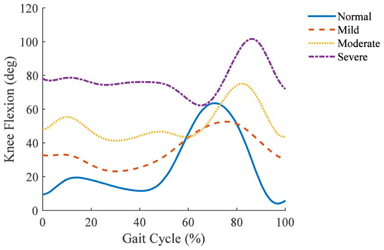
Patella alta is common in cerebral palsy, especially in patients with crouch gait. Correction of patella alta has been advocated in the treatment of crouch, however the appropriate degree of correction and the implications for knee extensor function remain unclear. Therefore, the goal of this study was to assess the impact of patellar position on quadriceps and patellar tendon forces during normal and crouch gait. To this end, a lower extremity musculoskeletal model with a novel 12 degree of freedom knee joint was used to simulate normal gait in a healthy child, as well as mild (23 deg min knee flexion in stance), moderate (41 deg), and severe (67 deg) crouch gait in three children with cerebral palsy. The simulations revealed that quadriceps and patellar tendon forces increase dramatically with crouch, and are modulated by patellar position. For example with a normal patellar tendon position, peak patellar tendon forces were 0.7 times body weight in normal walking, but reached 2.2, 3.2 and 5.4 times body weight in mild, moderate and severe crouch. Moderate patella alta acted to reduce quadriceps and patellar tendon loads in crouch gait, due to an enhancement of the patellar tendon moment arms with alta in a flexed knee. In contrast, patella baja reduced the patellar tendon moment arm in a flexed knee and thus induced an increase in the patellar tendon loads needed to walk in crouch. Functionally, these results suggest that patella baja could also compromise knee extensor function for other flexed knee activities such as chair rise and stair climbing. The findings are important to consider when using surgical approaches for correcting patella alta in children who exhibit crouch gait patterns.

Keywords: Cerebral palsy; Knee extensor; Moment arm; Patella alta; Patella baja; Patella position.


Figures

Figure 1
Figure 1
Lower extremity musculoskeletal model with novel knee joint with 6 degrees of freedom at both the tibiofemoral and patellofemoral joints. Ligament, cartilage, and bony geometries were taken from MR images, and the behavior was validated against dynamic MRI (Lenhart et al., 2015a).
Figure 2
Figure 2
Demonstration of the range of patellar positions used during gait simulations. Patella positions were classified as baja, normal, or alta based on the Insall-Salvati (IS) ratio (Insall and Salvati, 1971), (Normal: 0.8 < IS < 1.2). For ease of comparison, the inferior-superior patella position is also presented in centimeters (cm), with respect to the mean normal simulation (0.0 cm). These absolute positions are scaled to the size of the typically-developing child (height = 1.40 m) used for the normal gait simulations.
Figure 3
Figure 3
Knee flexion angle over the gait cycle for each of the subjects used in this analysis.
Figure 4
Figure 4
(Top) Quadriceps forces normalized to body weight (BW) over the stance phase of the gait cycle. Different colored lines indicate the different patellar positions simulated. Baja = Blue, Normal = Black, Alta = Red. (Bottom) Maximum quadriceps forces normalized by body weight for each patellar position. The shaded region is the normal range for patellar position. ** Simulations of severe crouch gait with the two extreme alta (IS = 1.92 and IS = 2.11) were not valid due to the lack of a wrapping constraint between the patellar tendon and distal femur. For large knee flexion angles, extreme alta caused unrealistic penetration of the patellar tendon within the distal femur.
Figure 5
Figure 5
(Top) Patellar tendon forces normalized to body weight (BW) over the stance phase of the gait cycle. Different colored lines indicate the different patellar positions simulated. Baja = Blue, Normal = Black, Alta = Red. (Middle) Maximum patellar tendon forces normalized by body weight for each patellar position. The shaded region is the normal range for patellar position. (Bottom) Patellar tendon moment arms (M.A) at the instant of maximal knee flexion during stance, for each patella position. Moment arms are scaled to the size of the typically-developing child (height = 1.40 m) used for the Normal Gait simulations. ** Simulations of severe crouch gait with the two extreme alta (IS = 1.92 and IS = 2.11) were not valid due to the lack of a wrapping constraint between the patellar tendon and distal femur. For large knee flexion angles, extreme alta caused unrealistic penetration of the patellar tendon within the distal femur.
Figure 6
Figure 6
Ratio of quadriceps to patellar tendon peak force during stance versus patellar position. Baja = Blue, Normal = Black, Alta = Red. The shaded region is the normal range for patellar position. ** Simulations of severe crouch gait with the two extreme alta (IS = 1.92 and IS = 2.11) were not valid due to the lack of a wrapping constraint between the patellar tendon and distal femur. For large knee flexion angles, extreme alta caused unrealistic penetration of the patellar tendon within the distal femur.
Figure 7
Figure 7
Demonstration of the interaction between patellar position and knee flexion angle on the magnitude and orientation of patellar contact force. Yellow arrow indicates the magnitude and direction of the patellar contact force acting on the femur. Force vectors are scaled for visualization; the scale factor is constant within each row, but differs between rows (crouch severity). Note that the quadriceps tendon wraps over the distal femur when baja is introduced in moderate and severe crouch, which contributes to the apparent decrease in the patello-femoral contact load in those cases.

References

    1. Arnold AS, Blemker SS, Delp SL. Evaluation of a deformable musculoskeletal model for estimating muscle-tendon lengths during crouch gait. Annals of Biomedical Engineering. 2001;29:263–274. - PubMed
    1. Arnold EM, Ward SR, Lieber RL, Delp SL. A model of the lower limb for analysis of human movement. Annals of Biomedical Engineering. 2010;38:269–279. - PMC - PubMed
    1. Askew M, Mow V. The biomechanical function of the collagen fibril ultrastructure of articular cartilage. Journal of biomechanical engineering. 1978;100:105–115.
    1. Beals RK. Treatment of knee contracture in cerebral palsy by hamstring lengthening, posterior capsulotomy, and quadriceps mechanism shortening. Dev Med Child Neurol. 2001;43:802–805. - PubMed
    1. Bei Y, Fregly BJ. Multibody dynamic simulation of knee contact mechanics. Medical engineering & physics. 2004;26:777–789. - PMC - PubMed