Sex specific recruitment of a medial prefrontal cortex-hippocampal-thalamic system during context-dependent renewal of responding to food cues in rats
- PMID: 27940080
- PMCID: PMC5334368
- DOI: 10.1016/j.nlm.2016.12.004
Sex specific recruitment of a medial prefrontal cortex-hippocampal-thalamic system during context-dependent renewal of responding to food cues in rats
Abstract
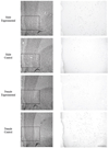
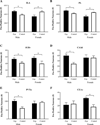
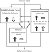
Renewal, or reinstatement, of responding to food cues after extinction may explain the inability to resist palatable foods and change maladaptive eating habits. Previously, we found sex differences in context-dependent renewal of extinguished Pavlovian conditioned responding to food cues. Context-induced renewal involves cue-food conditioning and extinction in different contexts and the renewal of conditioned behavior is induced by return to the conditioning context (ABA renewal). Male rats showed renewal of responding while females did not. In the current study we sought to identify recruitment of key neural systems underlying context-mediated renewal and sex differences. We examined Fos induction within the ventromedial prefrontal cortex (vmPFC), hippocampal formation, thalamus and amygdala in male and female rats during the test for renewal. We found sex differences in vmPFC recruitment during renewal. Male rats in the experimental condition showed renewal of responding and had more Fos induction within the infralimbic and prelimbic vmPFC areas compared to controls that remained in the same context throughout training and testing. Females in the experimental condition did not show renewal or an increase in Fos induction. Additionally, Fos expression differed between experimental and control groups and between the sexes in the hippocampal formation, thalamus and amygdala. Within the ventral subiculum, the experimental groups of both sexes had more Fos compared to control groups. Within the dorsal CA1 and the anterior region of the paraventricular nucleus of the thalamus, in males, the experimental group had higher Fos induction, while both females groups had similar number of Fos-positive neurons. Within the capsular part of the central amygdalar nucleus, females in the experimental group had higher Fos induction, while males groups had similar amounts. The differential recruitment corresponded to the behavioral differences between males and females and suggests the medial prefrontal cortex-hippocampal-thalamic system is a critical site of sex differences during renewal of appetitive Pavlovian responding to food cues. These findings provide evidence for novel neural mechanisms underlying sex differences in food motivation and contextual processing in associative learning and memory. The results should also inform future molecular and translational work investigating sex differences and maladaptive eating habits.
Keywords: Appetitive; Conditioning; Context; Medial prefrontal cortex; Renewal.
Copyright © 2016 Elsevier Inc. All rights reserved.
Figures


Similar articles
-
Distinct recruitment of the hippocampal, thalamic, and amygdalar neurons projecting to the prelimbic cortex in male and female rats during context-mediated renewal of responding to food cues.Neurobiol Learn Mem. 2018 Apr;150:25-35. doi: 10.1016/j.nlm.2018.02.013. Epub 2018 Feb 26. Neurobiol Learn Mem. 2018. PMID: 29496643 Free PMC article.
-
Renewal of conditioned responding to food cues in rats: Sex differences and relevance of estradiol.Physiol Behav. 2015 Nov 1;151:338-44. doi: 10.1016/j.physbeh.2015.07.035. Epub 2015 Aug 4. Physiol Behav. 2015. PMID: 26253218 Free PMC article.
-
Medial prefrontal cortex involvement in the expression of extinction and ABA renewal of instrumental behavior for a food reinforcer.Neurobiol Learn Mem. 2016 Feb;128:33-9. doi: 10.1016/j.nlm.2015.12.003. Epub 2015 Dec 23. Neurobiol Learn Mem. 2016. PMID: 26723281 Free PMC article.
-
Characterisation of the neural basis underlying appetitive extinction & renewal in Cacna1c rats.Neuropharmacology. 2023 Apr 1;227:109444. doi: 10.1016/j.neuropharm.2023.109444. Epub 2023 Jan 29. Neuropharmacology. 2023. PMID: 36724867 Review.
-
BEHAVIORAL AND NEUROBIOLOGICAL MECHANISMS OF PAVLOVIAN AND INSTRUMENTAL EXTINCTION LEARNING.Physiol Rev. 2021 Apr 1;101(2):611-681. doi: 10.1152/physrev.00016.2020. Epub 2020 Sep 24. Physiol Rev. 2021. PMID: 32970967 Free PMC article. Review.
Cited by
-
Distinct recruitment of the hippocampal, thalamic, and amygdalar neurons projecting to the prelimbic cortex in male and female rats during context-mediated renewal of responding to food cues.Neurobiol Learn Mem. 2018 Apr;150:25-35. doi: 10.1016/j.nlm.2018.02.013. Epub 2018 Feb 26. Neurobiol Learn Mem. 2018. PMID: 29496643 Free PMC article.
-
Sex differences in the immediate extinction deficit and renewal of extinguished fear in rats.PLoS One. 2022 Jun 10;17(6):e0264797. doi: 10.1371/journal.pone.0264797. eCollection 2022. PLoS One. 2022. PMID: 35687598 Free PMC article.
-
The Function of Paraventricular Thalamic Circuitry in Adaptive Control of Feeding Behavior.Front Behav Neurosci. 2021 Apr 27;15:671096. doi: 10.3389/fnbeh.2021.671096. eCollection 2021. Front Behav Neurosci. 2021. PMID: 33986649 Free PMC article.
-
Estrous cycle state-dependent renewal of appetitive behavior recruits unique patterns of Arc mRNA in female rats.Front Behav Neurosci. 2023 Jul 13;17:1210631. doi: 10.3389/fnbeh.2023.1210631. eCollection 2023. Front Behav Neurosci. 2023. PMID: 37521726 Free PMC article.
-
Persistent disruption of overexpectation learning after inactivation of the lateral orbitofrontal cortex in male rats.Psychopharmacology (Berl). 2023 Mar;240(3):501-511. doi: 10.1007/s00213-022-06198-2. Epub 2022 Aug 6. Psychopharmacology (Berl). 2023. PMID: 35932299
References
-
- Bhatnagar S, Dallman MF. The paraventricular nucleus of the thalamus alters rhythms in core temperature and energy balance in a state-dependent manner. Brain Res. 1999;851:66–75. - PubMed
MeSH terms
Substances
Grants and funding
LinkOut - more resources
Full Text Sources
Other Literature Sources
Miscellaneous

