. 2017 Mar 10:604:9-21.
doi: 10.1016/j.gene.2016.11.043. Epub 2016 Dec 6.

Cloning and functional characterizations of an apoptogenic Hid gene in the Scuttle Fly, Megaselia scalaris (Diptera; Phoridae)

Affiliations

Cloning and functional characterizations of an apoptogenic Hid gene in the Scuttle Fly, Megaselia scalaris (Diptera; Phoridae)

Siuk Yoo et al. Gene. .

Abstract

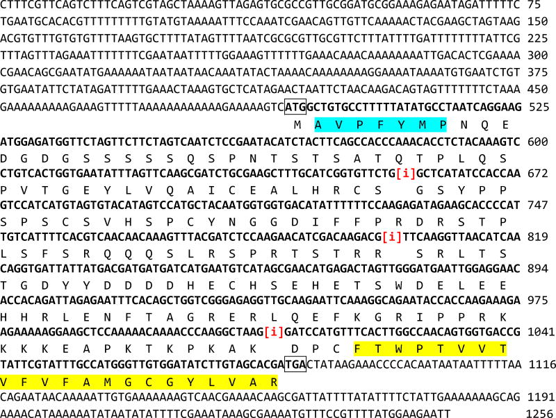
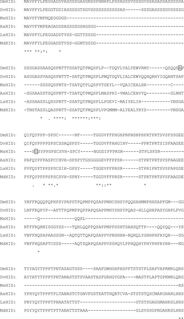
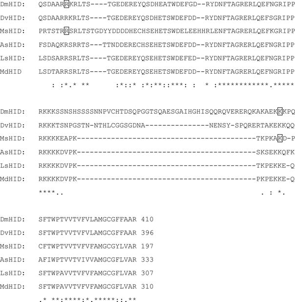
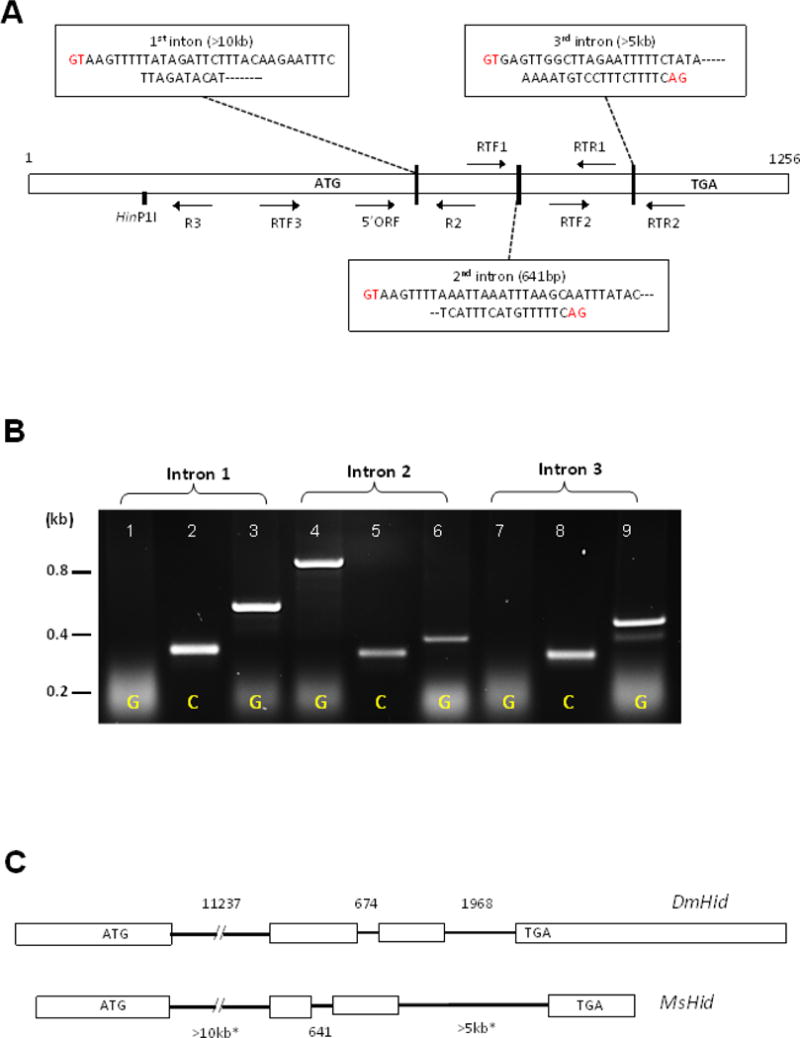
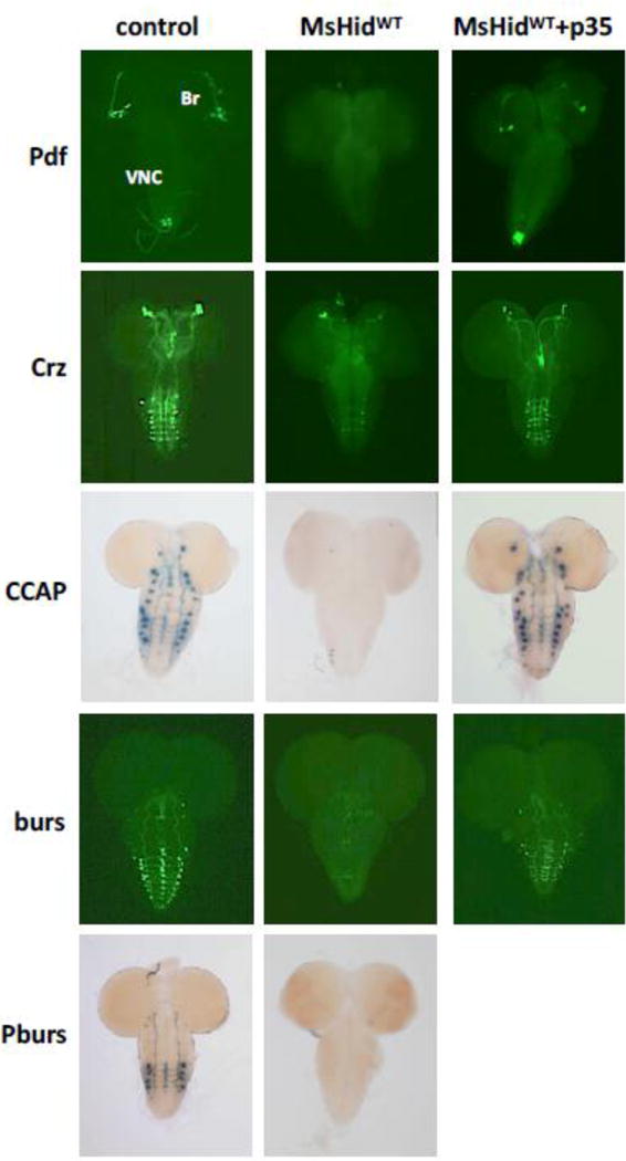
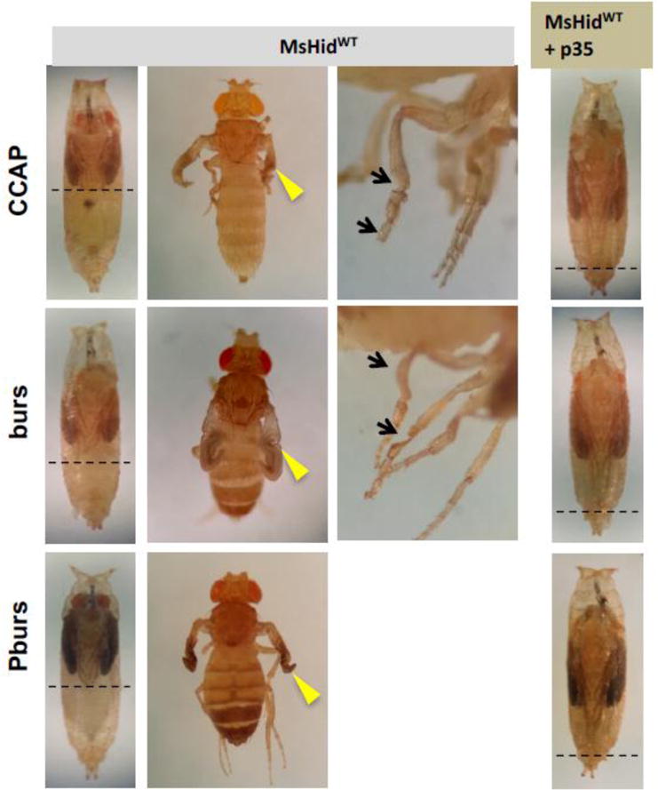
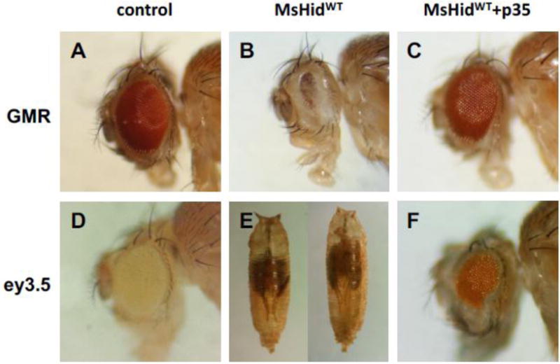
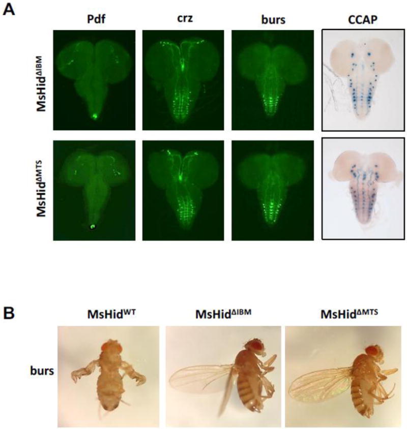
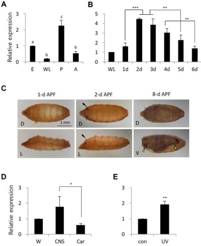
Although the mechanisms of apoptotic cell death have been well studied in the fruit fly, Drosophila melanogaster, it is unclear whether such mechanisms are conserved in other distantly related species. Using degenerate primers and PCR, we cloned a proapoptotic gene homologous to Head involution defective (Hid) from the Scuttle fly, Megaselia scalaris (MsHid). MsHid cDNA encodes a 197-amino acid-long polypeptide, which so far is the smallest HID protein. PCR analyses revealed that the MsHid gene consists of four exons and three introns. Ectopic expression of MsHid in various peptidergic neurons and non-neuronal tissues in Drosophila effectively induced apoptosis of these cells. However, deletion of either conserved domain, N-terminal IBM or C-terminal MTS, abolished the apoptogenic activity of MsHID, indicating that these two domains are indispensable. Expression of MsHid was found in all life stages, but more prominently in embryos and pupae. MsHid is actively expressed in the central nervous system (CNS), indicating its important role in CNS development. Together MsHID is likely to be an important cell death inducer during embryonic and post-embryonic development in this species. In addition, we found 2-fold induction of MsHid expression in UV-irradiated embryos, indicating a possible role for MsHid in UV-induced apoptosis.

Keywords: Apoptosis; Evolution; Hid; Peptidergic neurons.


Figures

Fig. 1
Fig. 1
Nucleotide sequence of the full-length MsHid cDNA. Open reading frame (ORF) is in bold-faced with start and stop codons in box. The ORF is predicted to encode 197-aa polypeptide. The positions of introns are indicated by [i]. Partial sequences of the introns are shown in Fig. 3. IBM domain is highlighted in blue, and MTS domain in yellow. These domains were subjected to site-directed mutagenesis.
Fig. 2
Fig. 2
Sequence alignment of the HID-homologous proteins. Identical residues are indicated by asterisks, and homologous residues by colons. Numbers indicate polypeptide length. Putative phosphorylation sites (Pro-X-Ser/Thr-Pro) by MAP kinase are highlighted. Residues in boxes in DmHID and MsHID indicate positions of introns. [Dv, D. virilis; Dm, D. melanogaster (Family Drosophilidae); As, Anastrepha suspensa (Family Tephritidae); Ms, Megaselia scalaris (Family Phoridae); Ls, Lucilia sericata (Family Calliphoridae); Md, Musca domestica (Family Muscidae)]. Phoridae is the only non-schizophoran family.
Fig. 2
Fig. 2
Sequence alignment of the HID-homologous proteins. Identical residues are indicated by asterisks, and homologous residues by colons. Numbers indicate polypeptide length. Putative phosphorylation sites (Pro-X-Ser/Thr-Pro) by MAP kinase are highlighted. Residues in boxes in DmHID and MsHID indicate positions of introns. [Dv, D. virilis; Dm, D. melanogaster (Family Drosophilidae); As, Anastrepha suspensa (Family Tephritidae); Ms, Megaselia scalaris (Family Phoridae); Ls, Lucilia sericata (Family Calliphoridae); Md, Musca domestica (Family Muscidae)]. Phoridae is the only non-schizophoran family.
Fig. 3
Fig. 3
Genomic organization of MsHid. A. Schematic diagram of exon-intron structure. The horizontal boxes indicate exons (1st exon, 657 bp; 2nd exon, 145 bp; 3rd exon, 205 bp; 4th exon 249 bp). The locations of three introns are indicated by vertical lines that are accompanied with partial sequences (consensus 5′ and 3′ splicing junctions are indicated in reds). Arrows indicate PCR primers. A HinP1 I site was used for inverse PCR. B. An agarose gel electrophoresis confirming introns. Primer sets are as follows: lane 1 and 2, 5′ORF-R2; lane 3, F3-Int1R; lane 4 and 5, RTF1-R1; lane 6, RTF1 and Int2R; lane 7 and 8, RTF2 and RTR2; lane 9, RTF2 and Int3R. Note that there is no PCR fragment in lane 1 and 7 due to large size of the 1st and the 3rd introns, respectively. However, PCR fragments from genomic DNA using the intron-derived primers match expected sizes (lane 3, 6, 9). (abbr.: G, genomic DNA template; C, cDNA template). C. Comparison of gene structure between DmHid (top) and MsHid (bottom). Exons are indicated by boxes, introns by lines. Numbers indicate nucleotide lengths of introns (asterisks for approximate values).
Fig. 4
Fig. 4
Apoptogenic activity of MsHid on indicated peptidergic neurons in the larval CNS. UAS-MsHidWT or UAS-MsHidWT; UAS-p35 were crossed with various Gal4 lines, each of which was combined with UAS-mCD8GFP (for Pdf, Crz, and burs) or UAS-lacZ reporter (for CCAP and Pburs). Ectopic expression of MsHid ablated almost all neuronal cells, whereas co-expression of p35 rescued the death. (Br, brain; VNC, ventral nerve cord)
Fig. 5
Fig. 5
Abnormal metamorphic development by the ablation of CCAP, burs, and Pburs neurons. UAS-MsHidWT or UAS-MsHidWT; UAS-p35 were crossed with indicated Gal4 lines. In all three cases, adult structures are abnormally formed in the pupal case (broken lines indicate posterior margin of the legs). Adult escapers show unexpanded wings (arrowheads) and shortened legs with crooked femurs and tibia, missing or dented tarsal segments (arrows). Co-expression of p35 restored normal pupal development.
Fig. 6
Fig. 6
Effect of ectopic expression of MsHid on the compound eyes. A–C. longGMR-Gal4 driven expression of MsHidWT (B) or MsHidWT + p35 (C). Severe degeneration of ommatidia is induced by MsHIDWT-mediated retinal cell death, but rescued by p35. D–F. Ectopic expression of MsHidWT by ey3.5-Gal4 resulted in lethality at pupal stage (E). Co-expression of p35 increase the survival rate up to 80%, but approximately one third of adult flies show partial degeneration (F) as compared to the control (D).
Fig. 7
Fig. 7
Role of IBM and MTS domains. A. Expression of UAS-MsHidΔIBM or UAS-MsHidΔMTS in four different peptidergic neurons. No sign of cell death was observed, except for CCAP-ΔIBM in which staining intensity is slightly lower in the neurons particularly in thoracic and subesophageal segments. B. Adult morphology. Expression of either mutant MsHid in burs neurons does not influence adult phenotype, as compared to unexpanded wings in MsHidWT expression.
Fig. 8
Fig. 8
Expression analysis of MsHid by real-time qPCR. Each bar indicates mean (± s.d.) of three independent experiments. A. Expression of MsHid during development (E-embryos; WL-wandering larvae; P-pupae; A-adults). Values are relative to those in embryos. Statistically different values are indicated by dissimilar letters (P<0.05 or smaller). B. MsHid expression during early metamorphosis. RNAs were prepared from wandering larvae (WL) and pupae from 1 to 6 days (d) APF. Values are relative to those in WL. C. Post-puparium development at 25°C. Within 1-d APF the puparium becomes tanned to brown (left panels), and the respiratory horns appear between 1- and 2-d APF (arrows in middle panels). At 8-d APF, adult structures (eyes and legs) are clearly visible (arrowhead in low-right panel). (D, dorsal view; L, lateral view; V, ventral view. In all images, anterior is to the left, and in lateral views, dorsal is up.). D. Relative expression levels of MsHid in the wandering larval stage. (W-whole larvae; CNS-central nervous system; Car-carcass). MsHid expression in the CNS is at least twice as much as that in the rest of the body. E. Two-fold expression of MsHid in response to UV irradiation. For B, D and E, significant differences were indicated by asterisks (*P<0.05; **P<0.01; ***P<0.001).

Similar articles

Cited by

References

    1. Bergmann A, Agapite J, McCall KA, Steller H. Drosophila gene hid is a direct molecular target of Ras-dependent survival signaling. Cell. 1998;95:331–341. - PubMed
    1. Bergmann A, Tugentman M, Shilo BZ, Steller H. Regulation of cell number by MAPK-dependent control of apoptosis: a mechanism for trophic survival signaling. Dev Cell. 2002;2:159–170. - PubMed
    1. Bilak A, Su TT. Regulation of Drosophila melanogaster pro-apoptotic gene hid. Apoptosis. 2009;14:943–949. - PMC - PubMed
    1. Brennecke J, Hipfner DR, Stark A, Russell RB, Cohen SM. bantam encodes a developmentally regulated microRNA that controls cell proliferation and regulates the proapoptotic gene hid in Drosophila. Cell. 2003;113:25–36. - PubMed
    1. Bryant B, Blair CD, Olson KE, Clem RJ. Annotation and expression profiling of apoptosis-related genes in the yellow fever mosquito, Aedes aegypti. Insect Biochem Mol Biol. 2008;38:331–345. - PMC - PubMed

MeSH terms

LinkOut - more resources