

Aquaporins in the Spinal Cord
- PMID: 27941618
- PMCID: PMC5187850
- DOI: 10.3390/ijms17122050
Aquaporins in the Spinal Cord
Abstract
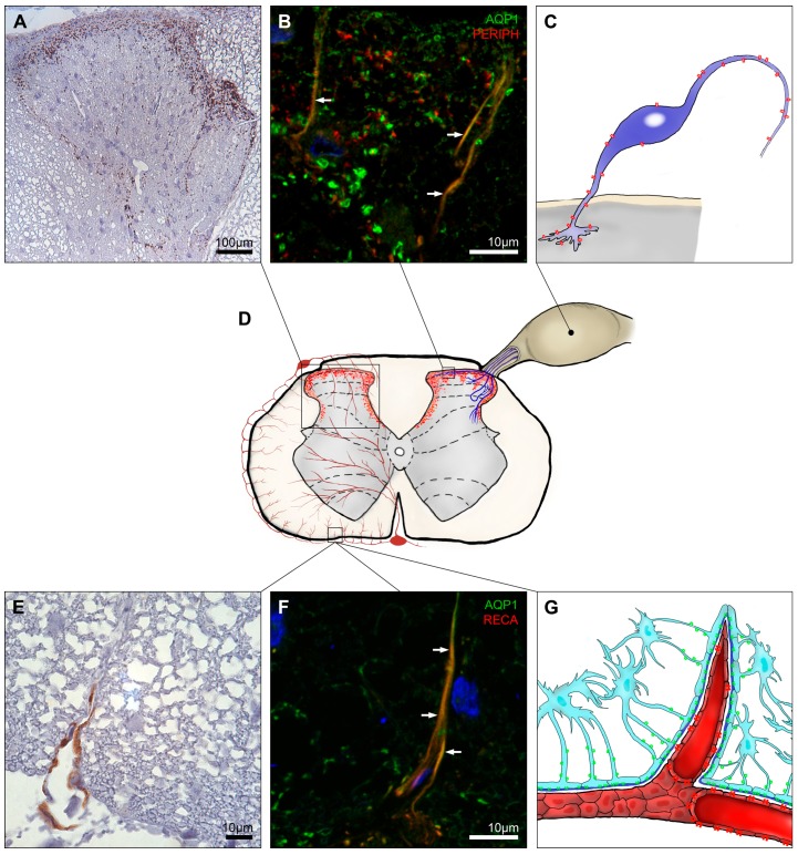
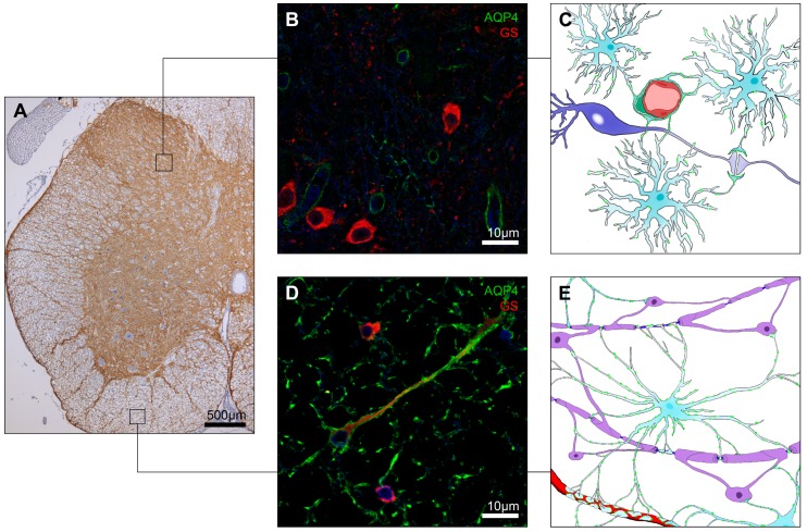
Aquaporins (AQPs) are water channel proteins robustly expressed in the central nervous system (CNS). A number of previous studies described the cellular expression sites and investigated their major roles and function in the brain and spinal cord. Among thirteen different mammalian AQPs, AQP1 and AQP4 have been mainly studied in the CNS and evidence has been presented that they play important roles in the pathogenesis of CNS injury, edema and multiple diseases such as multiple sclerosis, neuromyelitis optica spectrum disorders, amyotrophic lateral sclerosis, glioblastoma multiforme, Alzheimer's disease and Parkinson's disease. The objective of this review is to highlight the current knowledge about AQPs in the spinal cord and their proposed roles in pathophysiology and pathogenesis related to spinal cord lesions and injury.
Keywords: aquaporin; astrocytes; central nervous system; endothelial cells; neurons; peripheral nerve; spinal cord.
Conflict of interest statement
All authors declare no conflict of interest.
Figures
Similar articles
-
Immunolocalization of Water Channel Proteins AQP1 and AQP4 in Rat Spinal Cord.J Histochem Cytochem. 2014 Aug;62(8):598-611. doi: 10.1369/0022155414537495. Epub 2014 May 14. J Histochem Cytochem. 2014. PMID: 24828513
-
The role of aquaporin-4 in synaptic plasticity, memory and disease.Brain Res Bull. 2018 Jan;136:118-129. doi: 10.1016/j.brainresbull.2017.02.011. Epub 2017 Mar 6. Brain Res Bull. 2018. PMID: 28274814 Review.
-
Aquaporins in Nervous System.Adv Exp Med Biol. 2017;969:81-103. doi: 10.1007/978-94-024-1057-0_5. Adv Exp Med Biol. 2017. PMID: 28258567 Review.
-
Aquaporins in Nervous System.Adv Exp Med Biol. 2023;1398:99-124. doi: 10.1007/978-981-19-7415-1_7. Adv Exp Med Biol. 2023. PMID: 36717489
-
Expression of aquaporin water channels in mouse spinal cord.Neuroscience. 2004;127(3):685-93. doi: 10.1016/j.neuroscience.2004.03.016. Neuroscience. 2004. PMID: 15283967
Cited by
-
Ultra-High Field Diffusion MRI Reveals Early Axonal Pathology in Spinal Cord of ALS mice.Transl Neurodegener. 2018 Aug 8;7:20. doi: 10.1186/s40035-018-0122-z. eCollection 2018. Transl Neurodegener. 2018. PMID: 30128146 Free PMC article.
-
[Neuroprotective effects and mechanism of saikosaponin A on acute spinal cord injury in rats].Zhongguo Xiu Fu Chong Jian Wai Ke Za Zhi. 2017 Jul 15;31(7):825-829. doi: 10.7507/1002-1892.201702106. Zhongguo Xiu Fu Chong Jian Wai Ke Za Zhi. 2017. PMID: 29798527 Free PMC article. Chinese.
-
Pituitary Gonadotropins, Prolactin and Growth Hormone Differentially Regulate AQP1 Expression in the Porcine Ovarian Follicular Cells.Int J Mol Sci. 2017 Dec 21;19(1):5. doi: 10.3390/ijms19010005. Int J Mol Sci. 2017. PMID: 29267208 Free PMC article.
-
Expression, Distribution and Role of Aquaporins in Various Rhinologic Conditions.Int J Mol Sci. 2020 Aug 14;21(16):5853. doi: 10.3390/ijms21165853. Int J Mol Sci. 2020. PMID: 32824013 Free PMC article. Review.
-
Injury and mechanism of recombinant E. coli expressing STa on piglets colon.J Vet Med Sci. 2018 Feb 9;80(2):205-212. doi: 10.1292/jvms.17-0528. Epub 2017 Nov 29. J Vet Med Sci. 2018. PMID: 29187713 Free PMC article.
References
-
- Amiry-Moghaddam M., Otsuka T., Hurn P.D., Traystman R.J., Haug F.M., Froehner S.C., Adams M.E., Neely J.D., Agre P., Ottersen O.P., et al. An α-syntrophin-dependent pool of AQP4 in astroglial end-feet confers bidirectional water flow between blood and brain. Proc. Natl. Acad. Sci. USA. 2003;100:2106–2111. doi: 10.1073/pnas.0437946100. - DOI - PMC - PubMed
-
- Verkman A.S., Mitra A.K. Structure and function of aquaporin water channels. Am. J. Physiol. Ren. Physiol. 2000;278:F13–F28. - PubMed
Publication types
MeSH terms
Substances
LinkOut - more resources
Full Text Sources
Other Literature Sources
