

Evolution of the Cytolytic Pore-Forming Proteins (Actinoporins) in Sea Anemones
- PMID: 27941639
- PMCID: PMC5198562
- DOI: 10.3390/toxins8120368
Evolution of the Cytolytic Pore-Forming Proteins (Actinoporins) in Sea Anemones
Abstract
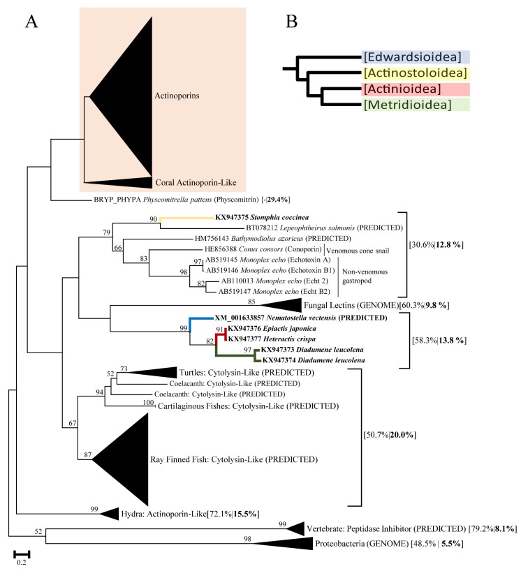
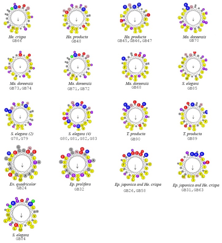
Sea anemones (Cnidaria, Anthozoa, and Actiniaria) use toxic peptides to incapacitate and immobilize prey and to deter potential predators. Their toxin arsenal is complex, targeting a variety of functionally important protein complexes and macromolecules involved in cellular homeostasis. Among these, actinoporins are one of the better characterized toxins; these venom proteins form a pore in cellular membranes containing sphingomyelin. We used a combined bioinformatic and phylogenetic approach to investigate how actinoporins have evolved across three superfamilies of sea anemones (Actinioidea, Metridioidea, and Actinostoloidea). Our analysis identified 90 candidate actinoporins across 20 species. We also found clusters of six actinoporin-like genes in five species of sea anemone (Nematostella vectensis, Stomphia coccinea, Epiactis japonica, Heteractis crispa, and Diadumene leucolena); these actinoporin-like sequences resembled actinoporins but have a higher sequence similarity with toxins from fungi, cone snails, and Hydra. Comparative analysis of the candidate actinoporins highlighted variable and conserved regions within actinoporins that may pertain to functional variation. Although multiple residues are involved in initiating sphingomyelin recognition and membrane binding, there is a high rate of replacement for a specific tryptophan with leucine (W112L) and other hydrophobic residues. Residues thought to be involved with oligomerization were variable, while those forming the phosphocholine (POC) binding site and the N-terminal region involved with cell membrane penetration were highly conserved.
Keywords: Cnidaria; cytolysins; target recognition; toxin.
Conflict of interest statement
The authors declare no conflict of interest. The funding agency had no role in the design of the research, in its performance, or in the interpretation of the results.
Figures


Similar articles
-
Multigene Family of Pore-Forming Toxins from Sea Anemone Heteractis crispa.Mar Drugs. 2018 May 24;16(6):183. doi: 10.3390/md16060183. Mar Drugs. 2018. PMID: 29794988 Free PMC article.
-
Identification of the two new, functional actinoporins, CJTOX I and CJTOX II, from the deep-sea anemone Cribrinopsis japonica.Toxicon. 2018 Jun 15;148:40-49. doi: 10.1016/j.toxicon.2018.04.008. Epub 2018 Apr 9. Toxicon. 2018. PMID: 29649486
-
Differential Effect of Membrane Composition on the Pore-Forming Ability of Four Different Sea Anemone Actinoporins.Biochemistry. 2016 Dec 6;55(48):6630-6641. doi: 10.1021/acs.biochem.6b01007. Epub 2016 Nov 22. Biochemistry. 2016. PMID: 27933793
-
The multigene families of actinoporins (part I): Isoforms and genetic structure.Toxicon. 2015 Sep;103:176-87. doi: 10.1016/j.toxicon.2015.06.028. Epub 2015 Jul 14. Toxicon. 2015. PMID: 26187849 Review.
-
Molecular mechanism of sphingomyelin-specific membrane binding and pore formation by actinoporins.Adv Exp Med Biol. 2010;677:106-15. Adv Exp Med Biol. 2010. PMID: 20687484 Review.
Cited by
-
New Insights into the Toxin Diversity and Antimicrobial Activity of the "Fire Coral" Millepora complanata.Toxins (Basel). 2022 Mar 14;14(3):206. doi: 10.3390/toxins14030206. Toxins (Basel). 2022. PMID: 35324703 Free PMC article.
-
Peptide Toxins from Antarctica: The Nemertean Predator and Scavenger Parborlasia corrugatus (McIntosh, 1876).Toxins (Basel). 2024 Apr 30;16(5):209. doi: 10.3390/toxins16050209. Toxins (Basel). 2024. PMID: 38787061 Free PMC article.
-
The Isolation of New Pore-Forming Toxins from the Sea Anemone Actinia fragacea Provides Insights into the Mechanisms of Actinoporin Evolution.Toxins (Basel). 2019 Jul 10;11(7):401. doi: 10.3390/toxins11070401. Toxins (Basel). 2019. PMID: 31295915 Free PMC article.
-
Characterising Functional Venom Profiles of Anthozoans and Medusozoans within Their Ecological Context.Mar Drugs. 2020 Apr 9;18(4):202. doi: 10.3390/md18040202. Mar Drugs. 2020. PMID: 32283847 Free PMC article. Review.
-
A Low Molecular Weight Protein from the Sea Anemone Anemonia viridis with an Anti-Angiogenic Activity.Mar Drugs. 2018 Apr 19;16(4):134. doi: 10.3390/md16040134. Mar Drugs. 2018. PMID: 29671760 Free PMC article.
References
Publication types
MeSH terms
Substances
LinkOut - more resources
Full Text Sources
Other Literature Sources
