Review
. 2016 Dec 9;17(12):2069.
doi: 10.3390/ijms17122069.

Glucose-6-Phosphate Dehydrogenase: Update and Analysis of New Mutations around the World

Affiliations
Review

Glucose-6-Phosphate Dehydrogenase: Update and Analysis of New Mutations around the World

Saúl Gómez-Manzo et al. Int J Mol Sci. .

Abstract

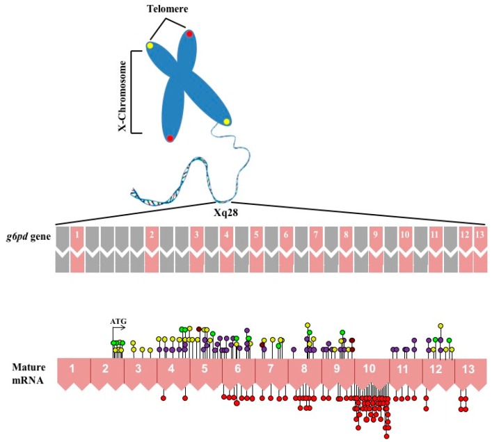
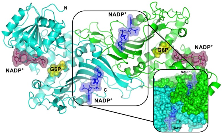
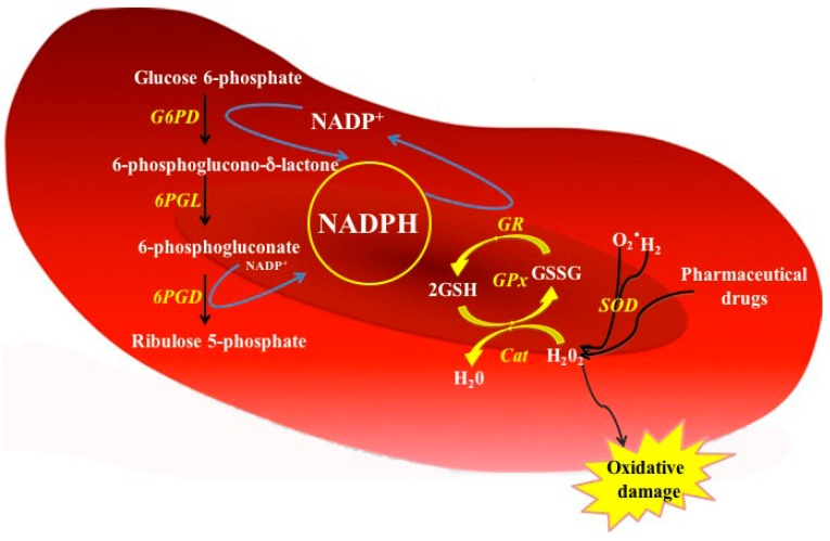
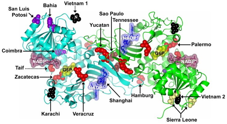
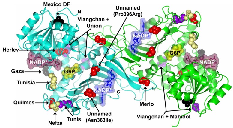
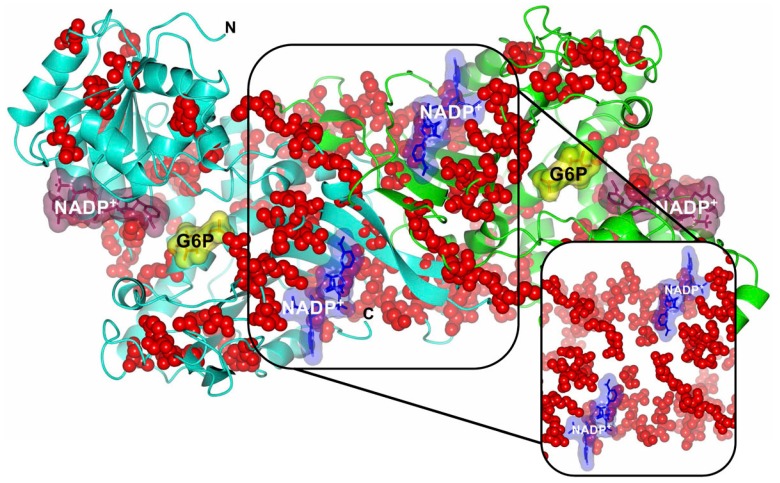
Glucose-6-phosphate dehydrogenase (G6PD) is a key regulatory enzyme in the pentose phosphate pathway which produces nicotinamide adenine dinucleotide phosphate (NADPH) to maintain an adequate reducing environment in the cells and is especially important in red blood cells (RBC). Given its central role in the regulation of redox state, it is understandable that mutations in the gene encoding G6PD can cause deficiency of the protein activity leading to clinical manifestations such as neonatal jaundice and acute hemolytic anemia. Recently, an extensive review has been published about variants in the g6pd gene; recognizing 186 mutations. In this work, we review the state of the art in G6PD deficiency, describing 217 mutations in the g6pd gene; we also compile information about 31 new mutations, 16 that were not recognized and 15 more that have recently been reported. In order to get a better picture of the effects of new described mutations in g6pd gene, we locate the point mutations in the solved three-dimensional structure of the human G6PD protein. We found that class I mutations have the most deleterious effects on the structure and stability of the protein.

Keywords: bioinformatics tools; clinical manifestations; glucose-6-phosphate dehydrogenase (G6PD) enzyme; mutations; three-dimensional structure.


Conflict of interest statement

The authors declare no conflict of interest.

Figures

Figure 1
Figure 1
Schematic overview of X chromosome and distribution of mutations in g6pd gene coding sequence. The top shown the introns and exons that are shown in gray and pink color boxes, respectively. The numbers (1–13) indicate exons of the human g6pd gene. In the bottom, the mRNA is schematized and all the single nucleotide substitutions (missense variants) are showed. The red circles are mutations associated with chronic nonspherocytic hemolytic anemia. Purple circles showed the Class II mutations. Class III mutations are shown in yellow circles. Class IV mutations are shown in brown circles and the unnamed reported class mutations are shown in bright green circles.
Figure 2
Figure 2
Crystallographic structure of the human wild-type (WT) G6PD enzyme (PDB entries 2BHL and 2BH9), showing the structural NADP+ (blue molecular surface), catalytic NADP+ (dark purple molecular surface), and G6P substrate (yellow molecular surface) in the dimer. The two monomers are shown in cyan and green. Right inset, close-up of the dimer interface and both structural NADP+ molecules. The figure was prepared using Collaborative Computational Project Number 4-Molecular Graphics (CCP4mg) (Didcot, UK) [28]. The same color code for G6PD enzyme is used in all other figures.
Figure 3
Figure 3
Function of G6PD enzyme in the PPP from red blood cells. In G6PD-normal red cells, the NADPH is produced by the action of glucose 6-phosphate dehydrogenase (G6PD) and 6-phosphogluconate dehydrogenase (6PGD) enzymes. The NADPH serves as proton donor to regenerates the GSSG oxidized. Cat = Catalase; GPx = Glutathione peroxidase; GR = Glutathione reductase; G6PD = glucose 6-phosphate dehydrogenase; 6PGL = 6-phosphogluconolactonase; 6GPD = 6-phosphogluconate dehydrogenase; SOD = Superoxide dismutase; GSH = Reduced glutathione; GSSG = Oxidized glutathione; H2O2 = Peroxide; O2 = Superoxide.
Figure 4
Figure 4
Mutations in the G6PD protein that have not been considered in the most recent review [34]. Structure of human WT G6PD enzyme (PDB entries 2BHL and 2BH9) indicating the location of Class I–IV mutations (missense variants) in the dimer (solid arrows). The Class I, II, III, and IV mutations are shown as red, purple, yellow, and brown spheres, respectively. The unnamed reported class mutations are shown as black spheres. Note that although the mutants are located on equivalent positions of G6PD dimer, Zacatecas, Palermo, Bahia, Vietnam 1 and 2, Sierra Leone, and San Luis Potosí mutants are shown in only one of the monomers. Also note that the Taif mutant is a deletion (dotted arrow) and Vietnam 1 mutant is represented by Val27 residue, since no electron density was observed for the 26 N-terminal residues where the Vietnam 1 mutant is located (dotted arrow). The figure was prepared using CCP4mg [28].
Figure 5
Figure 5
Recent described mutations in the G6PD protein. Structure of human WT G6PD enzyme (PDB entries 2BHL and 2BH9) indicating the location of Class I–IV mutations (missense variants) in the dimer. The Class I, II, III, and IV mutations are shown as red, purple, yellow, and brown spheres, respectively. The unnamed reported class mutations are shown as black spheres. Note that although most of the mutants are located on equivalent positions of G6PD dimer, Viangchan + Mahidol and Viangchan + Union mutants are shown in only one of the monomers. The figure was prepared using CCP4mg [28].
Figure 6
Figure 6
Structure of human WT G6PD enzyme (PDB entries 2BHL and 2BH9) indicating the location of Class I mutations in the dimer. Note that all mutations are located on equivalent positions of G6PD dimer. Right inset, close-up of Class I (red spheres) mutations located in both the dimer interface and near the structural NADP+ molecules. The figure was prepared using CCP4mg [28].

References

    1. Pai G.S., Sprenkle J.A., Do T.T., Mareni C.E., Migeon B.R. Localization of loci for hypoxanthine phosphoribosyltransferase and glucose-6-phosphate dehydrogenase 202 and biochemical evidence of nonrandom X chromosome expression from studies of a human X-autosome translocation. Proc. Natl. Acad. Sci. USA. 1980;77:2810–2813. doi: 10.1073/pnas.77.5.2810. - DOI - PMC - PubMed
    1. Luzzatto L., Battistuzzi G. Glucose-6-phosphate dehydrogenase. Adv. Hum. Genet. 1985;14:217–386. - PubMed
    1. Allahverdiyev A.M., Bagirova M., Elcicek S., Koc R.C., Ates S.C., Baydar S.Y., Yaman S., Abamor E.S., Oztel O.N. Glucose-6-Phosphate Dehydrogenase Deficiency and Malaria: A Method to Detect Primaquine-Induced Hemolysis In Vitro. In: Canuto R.A., editor. Biochemistry, Genetics and Molecular Biology. In Tech; Rijeka, Croatia: 2012.
    1. Szabo P., Purrello M., Rocchi M., Archidiacono N., Alhadeff B., Filippi G., Toniolo D., Martini G., Luzzatto L., Siniscalco M. Cytological mapping of the human glucose-6-phosphate dehydrogenase gene distal to the fragile-X site suggests a high rate of meiotic recombination across this site. Proc. Natl. Acad. Sci. USA. 1984;81:7855–7859. doi: 10.1073/pnas.81.24.7855. - DOI - PMC - PubMed
    1. Patterson M., Schwartz C., Bell M., Hofker M., Trask B., van den Engh G., Davies K.E. Physical mapping studies on the human X chromosome in the region Xq27-Xqter. Genomics. 1987;1:297–306. doi: 10.1016/0888-7543(87)90028-0. - DOI - PubMed

MeSH terms

Substances