Precise small-molecule recognition of a toxic CUG RNA repeat expansion
- PMID: 27941760
- PMCID: PMC5290590
- DOI: 10.1038/nchembio.2251
Precise small-molecule recognition of a toxic CUG RNA repeat expansion
Abstract
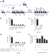
Excluding the ribosome and riboswitches, developing small molecules that selectively target RNA is a longstanding problem in chemical biology. A typical cellular RNA is difficult to target because it has little tertiary, but abundant secondary structure. We designed allele-selective compounds that target such an RNA, the toxic noncoding repeat expansion (r(CUG)exp) that causes myotonic dystrophy type 1 (DM1). We developed several strategies to generate allele-selective small molecules, including non-covalent binding, covalent binding, cleavage and on-site probe synthesis. Covalent binding and cleavage enabled target profiling in cells derived from individuals with DM1, showing precise recognition of r(CUG)exp. In the on-site probe synthesis approach, small molecules bound adjacent sites in r(CUG)exp and reacted to afford picomolar inhibitors via a proximity-based click reaction only in DM1-affected cells. We expanded this approach to image r(CUG)exp in its natural context.
Conflict of interest statement
The authors declare no competing financial interests.
Figures


Similar articles
-
Rationally designed small molecules that target both the DNA and RNA causing myotonic dystrophy type 1.J Am Chem Soc. 2015 Nov 11;137(44):14180-9. doi: 10.1021/jacs.5b09266. Epub 2015 Nov 3. J Am Chem Soc. 2015. PMID: 26473464
-
A CTG repeat-selective chemical screen identifies microtubule inhibitors as selective modulators of toxic CUG RNA levels.Proc Natl Acad Sci U S A. 2019 Oct 15;116(42):20991-21000. doi: 10.1073/pnas.1901893116. Epub 2019 Sep 30. Proc Natl Acad Sci U S A. 2019. PMID: 31570586 Free PMC article.
-
Precise small-molecule cleavage of an r(CUG) repeat expansion in a myotonic dystrophy mouse model.Proc Natl Acad Sci U S A. 2019 Apr 16;116(16):7799-7804. doi: 10.1073/pnas.1901484116. Epub 2019 Mar 29. Proc Natl Acad Sci U S A. 2019. PMID: 30926669 Free PMC article.
-
NMR structures of small molecules bound to a model of a CUG RNA repeat expansion.Bioorg Med Chem Lett. 2024 Oct 1;111:129888. doi: 10.1016/j.bmcl.2024.129888. Epub 2024 Jul 14. Bioorg Med Chem Lett. 2024. PMID: 39002937 Free PMC article. Review.
-
Gain of RNA function in pathological cases: Focus on myotonic dystrophy.Biochimie. 2011 Nov;93(11):2006-12. doi: 10.1016/j.biochi.2011.06.028. Epub 2011 Jul 13. Biochimie. 2011. PMID: 21763392 Review.
Cited by
-
The sustained expression of Cas9 targeting toxic RNAs reverses disease phenotypes in mouse models of myotonic dystrophy type 1.Nat Biomed Eng. 2021 Feb;5(2):157-168. doi: 10.1038/s41551-020-00607-7. Epub 2020 Sep 14. Nat Biomed Eng. 2021. PMID: 32929188 Free PMC article.
-
Understanding the characteristics of nonspecific binding of drug-like compounds to canonical stem-loop RNAs and their implications for functional cellular assays.RNA. 2021 Jan;27(1):12-26. doi: 10.1261/rna.076257.120. Epub 2020 Oct 7. RNA. 2021. PMID: 33028652 Free PMC article.
-
Surface-enhanced Raman spectroscopy for drug discovery: peptide-RNA binding.Anal Bioanal Chem. 2022 Aug;414(20):6009-6016. doi: 10.1007/s00216-022-04190-5. Epub 2022 Jun 29. Anal Bioanal Chem. 2022. PMID: 35764806 Free PMC article.
-
Targeting RNA with Small Molecules To Capture Opportunities at the Intersection of Chemistry, Biology, and Medicine.J Am Chem Soc. 2019 May 1;141(17):6776-6790. doi: 10.1021/jacs.8b13419. Epub 2019 Apr 19. J Am Chem Soc. 2019. PMID: 30896935 Free PMC article.
-
Hybrid splicing minigene and antisense oligonucleotides as efficient tools to determine functional protein/RNA interactions.Sci Rep. 2017 Dec 14;7(1):17587. doi: 10.1038/s41598-017-17816-x. Sci Rep. 2017. PMID: 29242583 Free PMC article.
References
-
- Schlünzen F, et al. Structural basis for the interaction of antibiotics with the peptidyl transferase center in eubacteria. Nature. 2001;413:814–821. - PubMed
-
- Carter AP, et al. Functional insights from the structure of the 30S ribosomal subunit and its interactions with antibiotics. Nature. 2000;407:340–348. - PubMed
-
- Howe JA, et al. Selective small-molecule inhibition of an RNA structural element. Nature. 2015;526:672–677. - PubMed
-
- Brook JD, et al. Molecular basis of myotonic dystrophy: expansion of a trinucleotide (CTG) repeat at the 3′ end of a transcript encoding a protein kinase family member. Cell. 1992;68:799–808. - PubMed
Publication types
MeSH terms
Substances
Associated data
- PubChem-Substance/318835077
- PubChem-Substance/318835080
- PubChem-Substance/318835081
- PubChem-Substance/318835082
- PubChem-Substance/318835083
- PubChem-Substance/318835084
- PubChem-Substance/318835085
- PubChem-Substance/318835086
- PubChem-Substance/318835087
- PubChem-Substance/318835088
- PubChem-Substance/318835089
- PubChem-Substance/318835090
- PubChem-Substance/318835078
- PubChem-Substance/318835079
Grants and funding
LinkOut - more resources
Full Text Sources
Other Literature Sources

