. 2016 Dec 12;5(12):e278.
doi: 10.1038/oncsis.2016.78.

In vivo functional dissection of a context-dependent role for Hif1α in pancreatic tumorigenesis

Affiliations

In vivo functional dissection of a context-dependent role for Hif1α in pancreatic tumorigenesis

T Cheng et al. Oncogenesis. .

Abstract

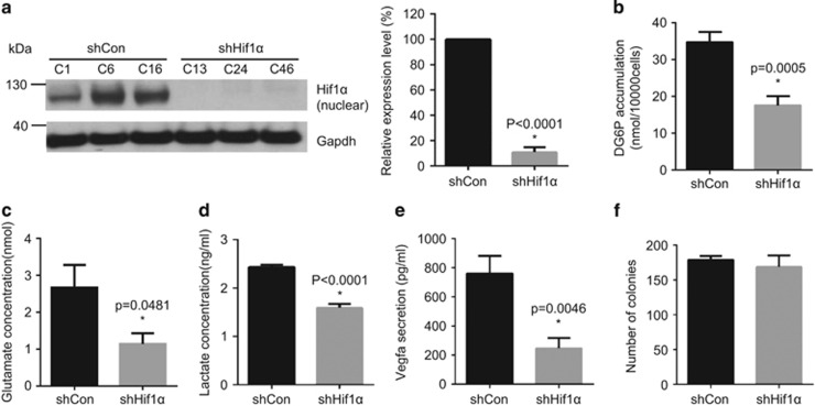
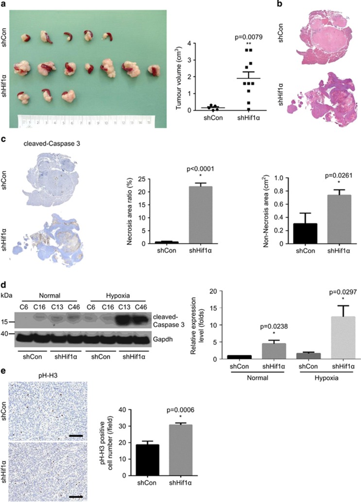
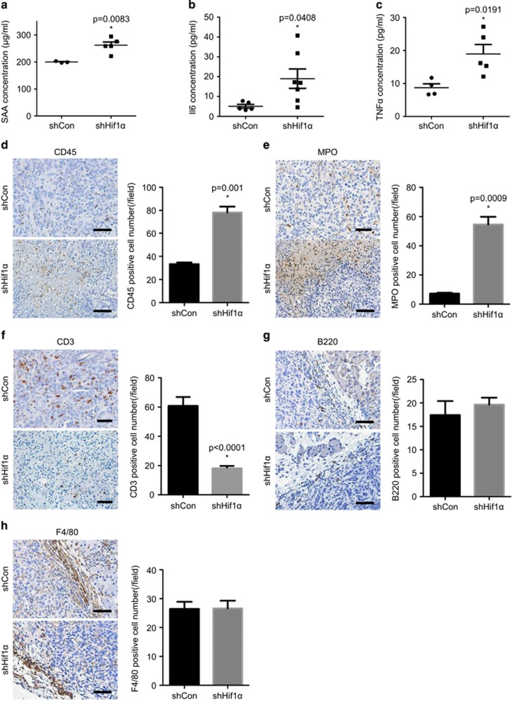
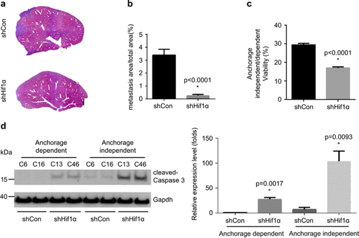
Hypoxia-inducible factor 1α (Hif1α) is a key regulator of cellular adaptation and survival under hypoxic conditions. In pancreatic ductal adenocarcinoma (PDAC), it has been recently shown that genetic ablation of Hif1α accelerates tumour development by promoting tumour-supportive inflammation in mice, questioning its role as the key downstream target of many oncogenic signals of PDAC. Likely, Hif1α has a context-dependent role in pancreatic tumorigenesis. To further analyse this, murine PDAC cell lines with reduced Hif1α expression were generated using shRNA transfection. Cells were transplanted into wild-type mice through orthotopic or portal vein injection in order to test the in vivo function of Hif1α in two major tumour-associated biological scenarios: primary tumour growth and remote colonization/metastasis. Although Hif1α protects PDAC cells from stress-induced cell deaths in both scenarios-in line with the general function Hif1α-its depletion leads to different oncogenic consequences. Hif1α depletion results in rapid tumour growth with marked hypoxia-induced cell death, which potentially leads to a persistent tumour-sustaining inflammatory response. However, it simultaneously reduces tumour colonization and hepatic metastases by increasing the susceptibility to anoikis induced by anchorage-independent conditions. Taken together, the role of Hif1α in pancreatic tumorigenesis is context-dependent. Clinical trials of Hif1α inhibitors need to take this into account, targeting the appropriate scenario, for example palliative vs adjuvant therapy.


Figures

Figure 1
Figure 1
Hif1α is downregulated in murine PDAC cells. (a) Western blot analyses show the downregulation of Hif1α in shHif1α cells but not in shControl cells: (left) western blot result; and (right) quantitative measurement; shHif1α: transfection with Hif1α-specific shRNA-expressing vector; shControl: transfection with control vectors. (b–d) The metabolic state of shControl cells or shHif1α cells is determined by glucose uptake assay (b), glutamate assay (c) and lactate secretion assay (d). (e) Vegfa secretion between shControl and shHif1α cells is measured by Vegfa ELISA assay. (f) The proliferation capacity of shControl or shHif1α cells is determined by colony formation assay. All data are presented as mean±s.e.m. (n=3), unpaired t-test is used to examine statistical significance, *P<0.05. See Supplementary Materials and Methods.
Figure 2
Figure 2
Hif1α depletion promotes primary tumour growth with tissue necrosis in vivo. (a) The orthotopic transplantation experiment shows the volume of tumours derived from shControl and shHif1α cells-injected animals: (left) gross pathology; and (right) quantitative measurement. (b) Representative H&E staining pictures show larger necrotic regions in shHif1α tumours compared with shControl tumours. (c) Immunohistochemistry (IHC) stainings of cleaved-caspase 3 (left) demonstrate more apoptotic cells (middle) and larger non-necrosis area (right) in shHif1α tumours in comparison with shControl tumours. (d) Western blot (left) and quantification results (right) show increased expression of cleaved-caspase 3 in shHif1α cells under hypoxic conditions. (e) IHC staining of phosph-histone H3 (pH-H3) and quantitative analysis demonstrates increased proliferation in shHif1α tumours; scale bar, 100 μm. See Supplementary Materials and Methods.
Figure 3
Figure 3
Increased inflammatory response triggered by Hif1α depletion. (a–c) Elevated levels of SAA (a), Il6 (b) and TNFα (c) are detected in the serums of shHif1α cells-transplanted animals compared with controls. (d) Representative IHC pictures of CD45 and quantitative analysis demonstrate increased immune cells infiltration (especially in necrotic areas) in the shHif1α tumours. (e) Representative IHC pictures of MPO and quantitative analysis reveal that neutrophils are the most infiltrated immune cells in the shHif1α tumours. (f) IHC staining of CD3 and quantitative analysis show reduced T-cell infiltration in the shHif1α tumours, which is confirmed by the quantitative analysis. Scale bar, 200 μm. (g–h) Representative IHC pictures and quantitative analysis of B220 (g) and F4/80 (h) show no difference in B cells and macrophages infiltration between shControl and shHif1α tumours. Scale bar, 200 μm. All data are presented as mean±s.e.m., and the statistical difference is determined by unpaired t-test. *P<0.05. See Supplementary Materials and Methods.
Figure 4
Figure 4
Loss of Hif1α impairs hepatic metastasis and resistance toward anoikis state. (a–b) Representative H&E staining pictures (a) and metastasis area calculation (b) reveal decreased metastatic foci after portal vein injection of shHif1α cells as compared with shControl cells. (c) Anoikis assay shows diminished cell survival in the shHif1α cells under anchorage-independent conditions. (d) Western blot and quantified measurements show expression of cleaved-caspase 3 in shControl or shHif1α cells under anchorage-dependent and -independent conditions. The data were a result of three independent experiment. An unpaired t-test was used for determining statistical significance. *P<0.05. See Supplementary Materials and Methods.

References

    1. Kong B, Cheng T, Wu W, Regel I, Raulefs S, Friess H et al. Hypoxia-induced endoplasmic reticulum stress characterizes a necrotic phenotype of pancreatic cancer. Oncotarget 2015; 31: 32154–32160. - PMC - PubMed
    1. Koong AC, Mehta VK, Le QT, Fisher GA, Terris DJ, Brown JM et al. Pancreatic tumors show high levels of hypoxia. Int J Radiat Oncol Biol Phys 2000; 48: 919–922. - PubMed
    1. Semenza GL. Regulation of mammalian O2 homeostasis by hypoxia-inducible factor 1. Annu Rev Cell Dev Biol 1999; 15: 551–578. - PubMed
    1. Kumar H, Choi DK. Hypoxia inducible factor pathway and physiological adaptation: a cell survival pathway? Mediators Inflamm 2015; 2015: 584758. - PMC - PubMed
    1. Colbert LE, Fisher SB, Balci S, Saka B, Chen Z, Kim S et al. High nuclear hypoxia-inducible factor 1 alpha expression is a predictor of distant recurrence in patients with resected pancreatic adenocarcinoma. Int J Radiat Oncol Biol Phys 2015; 91: 631–639. - PMC - PubMed

LinkOut - more resources