Overexpression of a Mycobacterium ulcerans Ag85B-EsxH Fusion Protein in Recombinant BCG Improves Experimental Buruli Ulcer Vaccine Efficacy
- PMID: 27941982
- PMCID: PMC5179062
- DOI: 10.1371/journal.pntd.0005229
Overexpression of a Mycobacterium ulcerans Ag85B-EsxH Fusion Protein in Recombinant BCG Improves Experimental Buruli Ulcer Vaccine Efficacy
Abstract
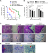
Buruli ulcer (BU) vaccine design faces similar challenges to those observed during development of prophylactic tuberculosis treatments. Multiple BU vaccine candidates, based upon Mycobacterium bovis BCG, altered Mycobacterium ulcerans (MU) cells, recombinant MU DNA, or MU protein prime-boosts, have shown promise by conferring transient protection to mice against the pathology of MU challenge. Recently, we have shown that a recombinant BCG vaccine expressing MU-Ag85A (BCG MU-Ag85A) displayed the highest level of protection to date, by significantly extending the survival time of MU challenged mice compared to BCG vaccination alone. Here we describe the generation, immunogenicity testing, and evaluation of protection conferred by a recombinant BCG strain which overexpresses a fusion of two alternative MU antigens, Ag85B and the MU ortholog of tuberculosis TB10.4, EsxH. Vaccination with BCG MU-Ag85B-EsxH induces proliferation of Ag85 specific CD4+ T cells in greater numbers than BCG or BCG MU-Ag85A and produces IFNγ+ splenocytes responsive to whole MU and recombinant antigens. In addition, anti-Ag85A and Ag85B IgG humoral responses are significantly enhanced after administration of the fusion vaccine compared to BCG or BCG MU-Ag85A. Finally, mice challenged with MU following a single subcutaneous vaccination with BCG MU-Ag85B-EsxH display significantly less bacterial burden at 6 and 12 weeks post-infection, reduced histopathological tissue damage, and significantly longer survival times compared to vaccination with either BCG or BCG MU-Ag85A. These results further support the potential of BCG as a foundation for BU vaccine design, whereby discovery and recombinant expression of novel immunogenic antigens could lead to greater anti-MU efficacy using this highly safe and ubiquitous vaccine.
Conflict of interest statement
The authors have declared that no competing interests exist.
Figures


Similar articles
-
Recombinant BCG Expressing Mycobacterium ulcerans Ag85A Imparts Enhanced Protection against Experimental Buruli ulcer.PLoS Negl Trop Dis. 2015 Sep 22;9(9):e0004046. doi: 10.1371/journal.pntd.0004046. eCollection 2015 Sep. PLoS Negl Trop Dis. 2015. PMID: 26393347 Free PMC article.
-
Analysis of the vaccine potential of plasmid DNA encoding nine mycolactone polyketide synthase domains in Mycobacterium ulcerans infected mice.PLoS Negl Trop Dis. 2014 Jan 2;8(1):e2604. doi: 10.1371/journal.pntd.0002604. eCollection 2014. PLoS Negl Trop Dis. 2014. PMID: 24392169 Free PMC article.
-
Prime-boost vaccination with Bacillus Calmette Guerin and a recombinant adenovirus co-expressing CFP10, ESAT6, Ag85A and Ag85B of Mycobacterium tuberculosis induces robust antigen-specific immune responses in mice.Mol Med Rep. 2015 Aug;12(2):3073-80. doi: 10.3892/mmr.2015.3770. Epub 2015 May 12. Mol Med Rep. 2015. PMID: 25962477
-
Buruli ulcer.Hum Vaccin. 2011 Nov;7(11):1198-203. doi: 10.4161/hv.7.11.17751. Epub 2011 Nov 1. Hum Vaccin. 2011. PMID: 22048117 Review.
-
[Novel vaccines against M. tuberculosis].Kekkaku. 2006 Dec;81(12):745-51. Kekkaku. 2006. PMID: 17240920 Review. Japanese.
Cited by
-
Unleashing the power of the BCG vaccine in modulating viral immunity through heterologous protection: A scoping review.Hum Vaccin Immunother. 2025 Dec;21(1):2521190. doi: 10.1080/21645515.2025.2521190. Epub 2025 Jul 3. Hum Vaccin Immunother. 2025. PMID: 40610004 Free PMC article.
-
Multi-epitope vaccine candidates based on mycobacterial membrane protein large (MmpL) proteins against Mycobacterium ulcerans.Open Biol. 2023 Nov;13(11):230330. doi: 10.1098/rsob.230330. Epub 2023 Nov 8. Open Biol. 2023. PMID: 37935359 Free PMC article.
-
In Silico Prediction of Epitopes in Virulence Proteins of Mycobacterium ulcerans for Vaccine Designing.Curr Genomics. 2021 Dec 31;22(7):512-525. doi: 10.2174/1389202922666211129113917. Curr Genomics. 2021. PMID: 35386432 Free PMC article.
-
The immunology of other mycobacteria: M. ulcerans, M. leprae.Semin Immunopathol. 2020 Jun;42(3):333-353. doi: 10.1007/s00281-020-00790-4. Epub 2020 Feb 25. Semin Immunopathol. 2020. PMID: 32100087 Free PMC article. Review.
-
Vaccine-Specific Immune Responses against Mycobacterium ulcerans Infection in a Low-Dose Murine Challenge Model.Infect Immun. 2020 Feb 20;88(3):e00753-19. doi: 10.1128/IAI.00753-19. Print 2020 Feb 20. Infect Immun. 2020. PMID: 31818964 Free PMC article.
References
-
- Nakanaga K, Yotsu RR, Hoshino Y, Suzuki K, Makino M, Ishii N. Buruli ulcer and mycolactone-producing mycobacteria. Japanese journal of infectious diseases. 2013;66(2):83–8. Epub 2013/03/22. - PubMed
-
- Organization WH. Buruli ulcer: progress report, 2004–2008. Releve epidemiologique hebdomadaire / Section d'hygiene du Secretariat de la Societe des Nations = Weekly epidemiological record / Health Section of the Secretariat of the League of Nations. 2008;83(17):145–54. Epub 2008/04/29.
Publication types
MeSH terms
Substances
LinkOut - more resources
Full Text Sources
Other Literature Sources
Research Materials

